 Movie
Movie Controller

[English] 日本語
 Yorodumi
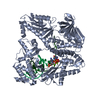
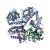
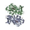
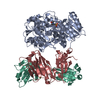
- PDB-1r5v: Evidence that structural rearrangements and/or flexibility during... -
+ Open data

- Basic information
| Entry | Database: PDB / ID: 1r5v | ||||||
|---|---|---|---|---|---|---|---|
| Title | Evidence that structural rearrangements and/or flexibility during TCR binding can contribute to T-cell activation | ||||||
 Components Components |
| ||||||
 Keywords Keywords |  SIGNALING PROTEIN / TCR / MHC class II / structural rearangement SIGNALING PROTEIN / TCR / MHC class II / structural rearangement | ||||||
| Function / homology |  Function and homology information Function and homology informationresponse to type II interferon / peptide antigen assembly with MHC class II protein complex / MHC class II protein complex / peptide antigen binding / antigen processing and presentation of exogenous peptide antigen via MHC class II / positive regulation of immune response / positive regulation of T cell activation / MHC class II protein complex binding / late endosome membrane / adaptive immune response ...response to type II interferon / peptide antigen assembly with MHC class II protein complex / MHC class II protein complex / peptide antigen binding / antigen processing and presentation of exogenous peptide antigen via MHC class II / positive regulation of immune response / positive regulation of T cell activation / MHC class II protein complex binding / late endosome membrane / adaptive immune response / membrane => GO:0016020 / lysosomal membraneSimilarity search - Function | ||||||
| Biological species |  Mus musculus (house mouse) Mus musculus (house mouse) | ||||||
| Method | X-RAY DIFFRACTION / SYNCHROTRON / MOLECULAR REPLACEMENT / Resolution: 2.5 Å | ||||||
 Authors Authors | Krogsgaard, M. / Prado, N. / Adams, E.J. / He, X.L. / Chow, D.C. / Wilson, D.B. / Garcia, K.C. / Davis, M.M. | ||||||
 Citation Citation | Journal: Mol.Cell / Year: 2003 Title: Evidence that structural rearrangements and/or flexibility during TCR binding can contribute to T cell activation. Authors: Krogsgaard, M. / Prado, N. / Adams, E.J. / He, X.L. / Chow, D.C. / Wilson, D.B. / Garcia, K.C. / Davis, M.M. | ||||||
| History |
|
- Structure visualization
| Structure viewer | Molecule: MolmilJmol/JSmol |
|---|
- Downloads & links
-Download
| PDBx/mmCIF format | 1r5v.cif.gz | 159.6 KB | Display | PDBx/mmCIF format |
|---|---|---|---|---|
| PDB format | pdb1r5v.ent.gz | 132.1 KB | Display | PDB format |
| PDBx/mmJSON format | 1r5v.json.gz | Tree view | PDBx/mmJSON format | |
| Others |  Other downloads Other downloads |
-Validation report
| Arichive directory | https://data.pdbj.org/pub/pdb/validation_reports/r5/1r5vftp://data.pdbj.org/pub/pdb/validation_reports/r5/1r5v | HTTPS FTP |
|---|
-Related structure data


-Links
 PDBj
PDBj


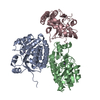
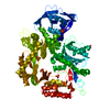
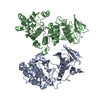
- Assembly
| Deposited unit | 
| ||||||||
|---|---|---|---|---|---|---|---|---|---|
| 1 |
| ||||||||
| 2 | 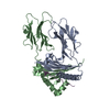
| ||||||||
| 3 | 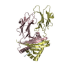
| ||||||||
| Unit cell |
|
-Components
| #1: Protein | Mass: 20962.383 Da / Num. of mol.: 2 Source method: isolated from a genetically manipulated source Source: (gene. exp.) Mus musculus (house mouse) / Plasmid: pGEMEX-1 / Production host:  Escherichia coli (E. coli) / Strain (production host): BL21(DE3)pLyss / References: UniProt: P04224 Escherichia coli (E. coli) / Strain (production host): BL21(DE3)pLyss / References: UniProt: P04224#2: Protein/peptide | Mass: 1410.656 Da / Num. of mol.: 2 / Source method: obtained synthetically / Details: artificial #3: Protein | Mass: 21737.195 Da / Num. of mol.: 2 Source method: isolated from a genetically manipulated source Source: (gene. exp.) Mus musculus (house mouse) / Plasmid: pGEMEX-1 / Production host: Escherichia coli (E. coli) / Strain (production host): BL21(DE3)pLyss / References: UniProt: P18468#4: Water | ChemComp-HOH / | Water Mass: 18.015 Da / Num. of mol.: 160 / Source method: isolated from a natural source / Formula: H2O Mass: 18.015 Da / Num. of mol.: 160 / Source method: isolated from a natural source / Formula: H2O |
|---|
-Experimental details
-Experiment
| Experiment | Method: X-RAY DIFFRACTION / Number of used crystals: 1 |
|---|
- Sample preparation
| Crystal | Density Matthews: 2.84 Å3/Da / Density % sol: 56.64 % | ||||||||||||||||||||||||||||||||||||||||||
|---|---|---|---|---|---|---|---|---|---|---|---|---|---|---|---|---|---|---|---|---|---|---|---|---|---|---|---|---|---|---|---|---|---|---|---|---|---|---|---|---|---|---|---|
| Crystal grow | Temperature: 298 K / Method: vapor diffusion, sitting drop / pH: 7 Details: PEG, pH 7, VAPOR DIFFUSION, SITTING DROP, temperature 298K | ||||||||||||||||||||||||||||||||||||||||||
| Crystal grow | *PLUS pH: 7.2 / Method: vapor diffusion, sitting drop | ||||||||||||||||||||||||||||||||||||||||||
| Components of the solutions | *PLUS
|
-Data collection
| Diffraction | Mean temperature: 100 K |
|---|---|
| Diffraction source | Source: SYNCHROTRON / Site: SSRL  / Beamline: BL11-1 / Wavelength: 1 Å / Beamline: BL11-1 / Wavelength: 1 Å |
| Detector | Type: ADSC QUANTUM 4 / Detector: CCD / Date: Nov 7, 2002 |
| Radiation | Protocol: SINGLE WAVELENGTH / Monochromatic (M) / Laue (L): M / Scattering type: x-ray |
| Radiation wavelength | Wavelength: 1 Å / Relative weight: 1 |
| Reflection | Resolution: 2.5→50 Å / Num. all: 34039 / Num. obs: 34039 / % possible obs: 94.4 % / Observed criterion σ(F): 0 / Observed criterion σ(I): 0 / Rmerge(I) obs: 0.063 / Net I/σ(I): 22.5 |
| Reflection shell | Resolution: 2.5→2.6 Å / Rmerge(I) obs: 0.377 / % possible all: 75.6 |
| Reflection | *PLUS Lowest resolution: 50 Å / Num. measured all: 144043 |
| Reflection shell | *PLUS % possible obs: 75.6 % / Mean I/σ(I) obs: 3.6 |
- Processing
| Software |
| ||||||||||||||||||||
|---|---|---|---|---|---|---|---|---|---|---|---|---|---|---|---|---|---|---|---|---|---|
| Refinement | Method to determine structure: MOLECULAR REPLACEMENT / Resolution: 2.5→50 Å / σ(F): 0 / Stereochemistry target values: Engh & Huber
| ||||||||||||||||||||
| Displacement parameters |
| ||||||||||||||||||||
| Refine analyze | Luzzati d res low obs: 6 Å | ||||||||||||||||||||
| Refinement step | Cycle: LAST / Resolution: 2.5→50 Å
| ||||||||||||||||||||
| Refine LS restraints |
| ||||||||||||||||||||
| LS refinement shell | Resolution: 2.5→2.6 Å / Rfactor Rfree: 0.294 / Rfactor Rwork: 0.284 | ||||||||||||||||||||
| Xplor file |
| ||||||||||||||||||||
| Refinement | *PLUS Lowest resolution: 50 Å / % reflection Rfree: 5 % | ||||||||||||||||||||
| Solvent computation | *PLUS | ||||||||||||||||||||
| Displacement parameters | *PLUS | ||||||||||||||||||||
| Refine LS restraints | *PLUS
|
