 Movie
Movie Controller


+ Open data

- Basic information
| Entry | Database: PDB / ID: 3l1q | |||||||||||||||||||||
|---|---|---|---|---|---|---|---|---|---|---|---|---|---|---|---|---|---|---|---|---|---|---|
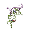
| Title | The crystal structure of the undecamer d(TGGCCTTAAGG) | |||||||||||||||||||||
 Components Components | 5'-D(* Keywords Keywords DNA / triple helix / triplet / Hoogsteen / reverse-Hoogsteen DNA / triple helix / triplet / Hoogsteen / reverse-HoogsteenFunction / homology | DNA / DNA (> 10) Function and homology information Function and homology informationMethod | X-RAY DIFFRACTION / SYNCHROTRON / MOLECULAR REPLACEMENT / Resolution: 2.5 Å  Authors AuthorsVan Hecke, K. |  CitationJournal: Cryst.Growth Des. / Year: 2010 CitationJournal: Cryst.Growth Des. / Year: 2010Title: Designing Triple Helical Fragments: The Crystal Structure of the Undecamer d(TGGCCTTAAGG) Mimicking T·AT Base Triplets Authors: Van Hecke, K. / Uytterhoeven, K. / Voet, A. / De Maeyer, M. / Van Meervelt, L. History |
|
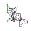
- Structure visualization
| Structure viewer | Molecule: MolmilJmol/JSmol |
|---|
- Downloads & links
-Download
| PDBx/mmCIF format | 3l1q.cif.gz | 22.6 KB | Display | PDBx/mmCIF format |
|---|---|---|---|---|
| PDB format | pdb3l1q.ent.gz | 14.8 KB | Display | PDB format |
| PDBx/mmJSON format | 3l1q.json.gz | Tree view | PDBx/mmJSON format | |
| Others |  Other downloads Other downloads |
-Validation report
| Arichive directory | https://data.pdbj.org/pub/pdb/validation_reports/l1/3l1qftp://data.pdbj.org/pub/pdb/validation_reports/l1/3l1q | HTTPS FTP |
|---|
-Related structure data
| Related structure data |  431dS S: Starting model for refinement |
|---|---|
| Similar structure data |


-Links
 PDBj
PDBj


- Assembly
| Deposited unit | 
| ||||||||
|---|---|---|---|---|---|---|---|---|---|
| 1 |
| ||||||||
| Unit cell |
|
-Components
| #1: DNA chain | Mass: 3389.221 Da / Num. of mol.: 2 / Source method: obtained synthetically Details: THE OLIGONUCLEOTIDE WAS PURCHASED FROM OSWEL DNA SERVICE (UNIVERSITY OF SOUTHAMPTON, UK) #2: Water | ChemComp-HOH / | Water Mass: 18.015 Da / Num. of mol.: 44 / Source method: isolated from a natural source / Formula: H2O Mass: 18.015 Da / Num. of mol.: 44 / Source method: isolated from a natural source / Formula: H2O |
|---|
-Experimental details
-Experiment
| Experiment | Method: X-RAY DIFFRACTION / Number of used crystals: 1 |
|---|
- Sample preparation
| Crystal | Density Matthews: 2.15 Å3/Da / Density % sol: 42.9 % |
|---|---|
| Crystal grow | Temperature: 289 K / Method: vapor diffusion, sitting drop / pH: 6 Details: 21.1mM sodium cacodylate, 47.4mM magnesium chloride, 21.1% MPD, pH 6.0, VAPOR DIFFUSION, SITTING DROP, temperature 289K |
-Data collection
| Diffraction | Mean temperature: 100 K |
|---|---|
| Diffraction source | Source: SYNCHROTRON / Site: EMBL/DESY, HAMBURG  / Beamline: X31 / Wavelength: 1.2 Å / Beamline: X31 / Wavelength: 1.2 Å |
| Detector | Type: MAR scanner 345 mm plate / Detector: IMAGE PLATE / Date: Apr 4, 2001 |
| Radiation | Monochromator: Si 111 horizontally focusing / Protocol: SINGLE WAVELENGTH / Monochromatic (M) / Laue (L): M / Scattering type: x-ray |
| Radiation wavelength | Wavelength: 1.2 Å / Relative weight: 1 |
| Reflection | Resolution: 2.5→32.4 Å / Num. obs: 2203 / % possible obs: 98.5 % / Redundancy: 3.1 % / Biso Wilson estimate: 86.8 Å2 / Rmerge(I) obs: 0.056 / Net I/σ(I): 14.9 |
| Reflection shell | Resolution: 2.5→2.64 Å / Redundancy: 3.2 % / Rmerge(I) obs: 0.583 / Mean I/σ(I) obs: 1.8 / Num. unique all: 307 / % possible all: 98.1 |
- Processing
| Software |
| |||||||||||||||||||||||||||||||||||
|---|---|---|---|---|---|---|---|---|---|---|---|---|---|---|---|---|---|---|---|---|---|---|---|---|---|---|---|---|---|---|---|---|---|---|---|---|
| Refinement | Method to determine structure: MOLECULAR REPLACEMENT Starting model: PDB ENTRY 431D Resolution: 2.5→20.76 Å / Cor.coef. Fo:Fc: 0.955 / Occupancy max: 1 / Occupancy min: 0.5 / SU B: 7.492 / SU ML: 0.171 / Isotropic thermal model: isotropic / σ(F): 0 / ESU R: 0.851 / Stereochemistry target values: Engh & Huber Details: MAXIMUM LIKELIHOOD; in the final refinement cycles of the structure, no Rfree was used. Please see the details in the primary citation.
| |||||||||||||||||||||||||||||||||||
| Solvent computation | Ion probe radii: 0.8 Å / Shrinkage radii: 0.8 Å / VDW probe radii: 1.4 Å / Solvent model: MASK | |||||||||||||||||||||||||||||||||||
| Displacement parameters | Biso max: 86.26 Å2 / Biso mean: 50.199 Å2 / Biso min: 28.89 Å2
| |||||||||||||||||||||||||||||||||||
| Refinement step | Cycle: LAST / Resolution: 2.5→20.76 Å
| |||||||||||||||||||||||||||||||||||
| Refine LS restraints |
| |||||||||||||||||||||||||||||||||||
| LS refinement shell | Resolution: 2.5→2.565 Å / Total num. of bins used: 20
|
