 Movie
Movie Controller

[English] 日本語
 Yorodumi
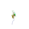
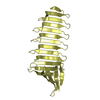
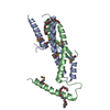
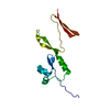
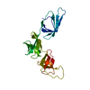
- PDB-3l1n: Crystal structure of Mp1p ligand binding domain 2 complexd with p... -
+ Open data

- Basic information
| Entry | Database: PDB / ID: 3l1n | ||||||
|---|---|---|---|---|---|---|---|
| Title | Crystal structure of Mp1p ligand binding domain 2 complexd with palmitic acid | ||||||
 Components Components | Cell wall antigen | ||||||
 Keywords Keywords | LIPID BINDING PROTEIN /  helix-turn-helix / protein-ligand complex helix-turn-helix / protein-ligand complex | ||||||
| Function / homology |  Function and homology information Function and homology informationyeast-form cell wall / structural constituent of cell wall / hyphal cell wall / fungal-type cell wall organization / fungal-type cell wall / fatty acid binding / extracellular regionSimilarity search - Function | ||||||
| Biological species |  Penicillium marneffei (fungus) Penicillium marneffei (fungus) | ||||||
| Method | X-RAY DIFFRACTION / SYNCHROTRON / SAD / Resolution: 1.3 Å | ||||||
 Authors Authors | Liao, S. / Tung, E.T. / Zheng, W. / Chong, K. / Xu, Y. / Bartlam, M. / Rao, Z. / Yuen, K.Y. | ||||||
 Citation Citation | Journal: J.Biol.Chem. / Year: 2010 Title: Crystal structure of the Mp1p ligand binding domain 2 reveals its function as a fatty acid-binding protein. Authors: Liao, S. / Tung, E.T. / Zheng, W. / Chong, K. / Xu, Y. / Dai, P. / Guo, Y. / Bartlam, M. / Yuen, K.Y. / Rao, Z. | ||||||
| History |
|
- Structure visualization
| Structure viewer | Molecule: MolmilJmol/JSmol |
|---|
- Downloads & links
-Download
| PDBx/mmCIF format | 3l1n.cif.gz | 79.4 KB | Display | PDBx/mmCIF format |
|---|---|---|---|---|
| PDB format | pdb3l1n.ent.gz | 63.5 KB | Display | PDB format |
| PDBx/mmJSON format | 3l1n.json.gz | Tree view | PDBx/mmJSON format | |
| Others |  Other downloads Other downloads |
-Validation report
| Arichive directory | https://data.pdbj.org/pub/pdb/validation_reports/l1/3l1nftp://data.pdbj.org/pub/pdb/validation_reports/l1/3l1n | HTTPS FTP |
|---|
-Related structure data
| Similar structure data |
|---|

-Links
 PDBj
PDBj


- Assembly
| Deposited unit | 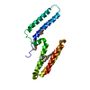
| ||||||||
|---|---|---|---|---|---|---|---|---|---|
| 1 |
| ||||||||
| 2 | 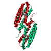
| ||||||||
| Unit cell |
| ||||||||
| Components on special symmetry positions |
|
-Components
| #1: Protein | Mass: 21204.037 Da / Num. of mol.: 1 / Fragment: ligand binding domain 2 / Mutation: I207M,L276M Source method: isolated from a genetically manipulated source Source: (gene. exp.) Penicillium marneffei (fungus) / Strain: PM4 / Gene: MP1 / Plasmid: pET28a / Production host:  Escherichia coli (E. coli) / Strain (production host): BL21 / References: UniProt: O42721 Escherichia coli (E. coli) / Strain (production host): BL21 / References: UniProt: O42721 |
|---|---|
| #2: Chemical | ChemComp-PLM / Palmitic acid  Mass: 256.424 Da / Num. of mol.: 1 / Source method: obtained synthetically / Formula: C16H32O2 Mass: 256.424 Da / Num. of mol.: 1 / Source method: obtained synthetically / Formula: C16H32O2 |
| #3: Water | ChemComp-HOH / Water Mass: 18.015 Da / Num. of mol.: 243 / Source method: isolated from a natural source / Formula: H2O Mass: 18.015 Da / Num. of mol.: 243 / Source method: isolated from a natural source / Formula: H2O |
-Experimental details
-Experiment
| Experiment | Method: X-RAY DIFFRACTION / Number of used crystals: 1 |
|---|
- Sample preparation
| Crystal | Density Matthews: 1.9 Å3/Da / Density % sol: 35.43 % |
|---|---|
| Crystal grow | Temperature: 289 K / Method: vapor diffusion, hanging drop / pH: 5.5 Details: 11% polyethylene glycol(PEG)8000, 0.1M HEPES(pH 7.5), 8% ethylene glycerol, VAPOR DIFFUSION, HANGING DROP, temperature 289K |
-Data collection
| Diffraction | Mean temperature: 100 K |
|---|---|
| Diffraction source | Source: SYNCHROTRON / Site: Photon Factory  / Beamline: BL-5A / Wavelength: 0.97894 Å / Beamline: BL-5A / Wavelength: 0.97894 Å |
| Detector | Type: ADSC QUANTUM 315 / Detector: CCD / Date: Jun 5, 2008 |
| Radiation | Protocol: SINGLE WAVELENGTH / Monochromatic (M) / Laue (L): M / Scattering type: x-ray |
| Radiation wavelength | Wavelength: 0.97894 Å / Relative weight: 1 |
| Reflection | Resolution: 1.3→19.74 Å / Num. all: 41553 / Num. obs: 40950 / % possible obs: 98 % / Observed criterion σ(F): 0 / Observed criterion σ(I): 0 / Redundancy: 20.2 % / Rmerge(I) obs: 0.079 |
| Reflection shell | Resolution: 1.3→1.35 Å / Redundancy: 16.2 % / Rmerge(I) obs: 0.383 / Mean I/σ(I) obs: 5.5 / Num. unique all: 4048 / % possible all: 95 |
- Processing
| Software |
| ||||||||||||||||||||||||||||||||||||||||||||||||||||||||||||||||||||||||||||||||||||||||||
|---|---|---|---|---|---|---|---|---|---|---|---|---|---|---|---|---|---|---|---|---|---|---|---|---|---|---|---|---|---|---|---|---|---|---|---|---|---|---|---|---|---|---|---|---|---|---|---|---|---|---|---|---|---|---|---|---|---|---|---|---|---|---|---|---|---|---|---|---|---|---|---|---|---|---|---|---|---|---|---|---|---|---|---|---|---|---|---|---|---|---|---|
| Refinement | Method to determine structure: SAD / Resolution: 1.3→19.74 Å / Cor.coef. Fo:Fc: 0.955 / Cor.coef. Fo:Fc free: 0.954 / SU B: 1.376 / SU ML: 0.028 / σ(F): 0 / ESU R: 0.059 / ESU R Free: 0.052 / Stereochemistry target values: Engh & Huber
| ||||||||||||||||||||||||||||||||||||||||||||||||||||||||||||||||||||||||||||||||||||||||||
| Solvent computation | Ion probe radii: 0.8 Å / Shrinkage radii: 0.8 Å / VDW probe radii: 1.2 Å / Solvent model: MASK | ||||||||||||||||||||||||||||||||||||||||||||||||||||||||||||||||||||||||||||||||||||||||||
| Displacement parameters | Biso mean: 14.547 Å2
| ||||||||||||||||||||||||||||||||||||||||||||||||||||||||||||||||||||||||||||||||||||||||||
| Refinement step | Cycle: LAST / Resolution: 1.3→19.74 Å
| ||||||||||||||||||||||||||||||||||||||||||||||||||||||||||||||||||||||||||||||||||||||||||
| Refine LS restraints |
| ||||||||||||||||||||||||||||||||||||||||||||||||||||||||||||||||||||||||||||||||||||||||||
| LS refinement shell | Resolution: 1.3→1.334 Å / Total num. of bins used: 20
|
