 Movie
Movie Controller


+ Open data

- Basic information
| Entry | Database: PDB / ID: 3emi | ||||||
|---|---|---|---|---|---|---|---|
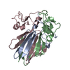
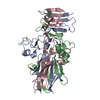
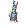
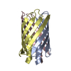
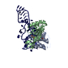
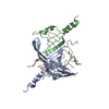
| Title | Crystal structure of Hia 307-422 non-adhesive domain | ||||||
 Components Components | Hia (Adhesin) | ||||||
 Keywords Keywords |  CELL ADHESION / non-adhesive domain / Hia adhesin / trimeric autotransporter / Haemophilus influenzae CELL ADHESION / non-adhesive domain / Hia adhesin / trimeric autotransporter / Haemophilus influenzae | ||||||
| Function / homology |  Function and homology information Function and homology information | ||||||
| Biological species |  Haemophilus influenzae (bacteria) Haemophilus influenzae (bacteria) | ||||||
| Method | X-RAY DIFFRACTION / SYNCHROTRON / MAD / Resolution: 1.8 Å | ||||||
 Authors Authors | Meng, G. / Waksman, G. | ||||||
 Citation Citation | Journal: J.Mol.Biol. / Year: 2008 Title: Repetitive Architecture of the Haemophilus influenzae Hia Trimeric Autotransporter Authors: Meng, G. / St Geme, J.W. / Waksman, G. | ||||||
| History |
|
- Structure visualization
| Structure viewer | Molecule: MolmilJmol/JSmol |
|---|
- Downloads & links
-Download
| PDBx/mmCIF format | 3emi.cif.gz | 56.5 KB | Display | PDBx/mmCIF format |
|---|---|---|---|---|
| PDB format | pdb3emi.ent.gz | 41.6 KB | Display | PDB format |
| PDBx/mmJSON format | 3emi.json.gz | Tree view | PDBx/mmJSON format | |
| Others |  Other downloads Other downloads |
-Validation report
| Arichive directory | https://data.pdbj.org/pub/pdb/validation_reports/em/3emiftp://data.pdbj.org/pub/pdb/validation_reports/em/3emi | HTTPS FTP |
|---|
-Related structure data


-Links
 PDBj
PDBj- Assembly
| Deposited unit | 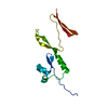
| ||||||||||||
|---|---|---|---|---|---|---|---|---|---|---|---|---|---|
| 1 | 
| ||||||||||||
| Unit cell |
| ||||||||||||
| Components on special symmetry positions |
|
-Components
| #1: Protein | Mass: 12047.246 Da / Num. of mol.: 1 / Fragment: UNP residues 307-422 / Mutation: I324M Source method: isolated from a genetically manipulated source Source: (gene. exp.) Haemophilus influenzae (bacteria) / Plasmid: pET15b / Production host: Escherichia coli (E. coli) / Strain (production host): BL21(B834) / References: UniProt: Q48152 |
|---|---|
| #2: Water | ChemComp-HOH / Water Mass: 18.015 Da / Num. of mol.: 244 / Source method: isolated from a natural source / Formula: H2O Mass: 18.015 Da / Num. of mol.: 244 / Source method: isolated from a natural source / Formula: H2O |
-Experimental details
-Experiment
| Experiment | Method: X-RAY DIFFRACTION / Number of used crystals: 1 |
|---|
- Sample preparation
| Crystal | Density Matthews: 2.64 Å3/Da / Density % sol: 53.34 % |
|---|---|
| Crystal grow | Temperature: 298 K / Method: vapor diffusion, hanging drop / Details: VAPOR DIFFUSION, HANGING DROP, temperature 298K |
-Data collection
| Diffraction source | Source: SYNCHROTRON / Site: ESRF  / Beamline: ID14-4 / Wavelength: 0.9785, 0.9790, 0.9750 / Beamline: ID14-4 / Wavelength: 0.9785, 0.9790, 0.9750 | ||||||||||||
|---|---|---|---|---|---|---|---|---|---|---|---|---|---|
| Detector | Type: ADSC QUANTUM 315 / Detector: CCD / Date: Nov 11, 2007 | ||||||||||||
| Radiation | Protocol: MAD / Monochromatic (M) / Laue (L): M / Scattering type: x-ray | ||||||||||||
| Radiation wavelength |
| ||||||||||||
| Reflection | Resolution: 1.8→30 Å / Num. obs: 12852 / % possible obs: 99.7 % / Observed criterion σ(F): 0 / Observed criterion σ(I): 0 / Redundancy: 32.6 % / Biso Wilson estimate: 15.127 Å2 / Rmerge(I) obs: 0.098 / Rsym value: 0.098 / Net I/σ(I): 42 | ||||||||||||
| Reflection shell | Resolution: 1.8→1.86 Å / Redundancy: 26 % / Rmerge(I) obs: 0.27 / Mean I/σ(I) obs: 11.3 / Rsym value: 0.27 / % possible all: 99.9 |
- Processing
| Software |
| ||||||||||||||||||||||||||||||||||||||||||||||||||||||||||||||||||||||||||||||||||||||||||||||||||||||||||||||||||||||||||||||||||||||||||||||||||||||
|---|---|---|---|---|---|---|---|---|---|---|---|---|---|---|---|---|---|---|---|---|---|---|---|---|---|---|---|---|---|---|---|---|---|---|---|---|---|---|---|---|---|---|---|---|---|---|---|---|---|---|---|---|---|---|---|---|---|---|---|---|---|---|---|---|---|---|---|---|---|---|---|---|---|---|---|---|---|---|---|---|---|---|---|---|---|---|---|---|---|---|---|---|---|---|---|---|---|---|---|---|---|---|---|---|---|---|---|---|---|---|---|---|---|---|---|---|---|---|---|---|---|---|---|---|---|---|---|---|---|---|---|---|---|---|---|---|---|---|---|---|---|---|---|---|---|---|---|---|---|---|---|
| Refinement | Method to determine structure: MAD / Resolution: 1.8→29.43 Å / Occupancy max: 1 / Occupancy min: 1 / FOM work R set: 0.878 / Isotropic thermal model: isotropic / Cross valid method: THROUGHOUT / Stereochemistry target values: Engh & Huber / Details: used weighted full matrix least squares procedure
| ||||||||||||||||||||||||||||||||||||||||||||||||||||||||||||||||||||||||||||||||||||||||||||||||||||||||||||||||||||||||||||||||||||||||||||||||||||||
| Solvent computation | Bsol: 54.433 Å2 / ksol: 0.328 e/Å3 | ||||||||||||||||||||||||||||||||||||||||||||||||||||||||||||||||||||||||||||||||||||||||||||||||||||||||||||||||||||||||||||||||||||||||||||||||||||||
| Displacement parameters | Biso max: 63.5 Å2 / Biso mean: 21.274 Å2 / Biso min: 5.5 Å2
| ||||||||||||||||||||||||||||||||||||||||||||||||||||||||||||||||||||||||||||||||||||||||||||||||||||||||||||||||||||||||||||||||||||||||||||||||||||||
| Refinement step | Cycle: LAST / Resolution: 1.8→29.43 Å
| ||||||||||||||||||||||||||||||||||||||||||||||||||||||||||||||||||||||||||||||||||||||||||||||||||||||||||||||||||||||||||||||||||||||||||||||||||||||
| Refine LS restraints |
| ||||||||||||||||||||||||||||||||||||||||||||||||||||||||||||||||||||||||||||||||||||||||||||||||||||||||||||||||||||||||||||||||||||||||||||||||||||||
| LS refinement shell |
|
