 Movie
Movie Controller

[English] 日本語
 Yorodumi
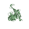
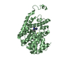
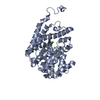
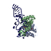
- PDB-2ccz: Crystal structure of E. coli primosomol protein PriB bound to ssDNA -
+ Open data

- Basic information
| Entry | Database: PDB / ID: 2ccz | ||||||
|---|---|---|---|---|---|---|---|
| Title | Crystal structure of E. coli primosomol protein PriB bound to ssDNA | ||||||
 Components Components |
| ||||||
 Keywords Keywords | DNA/REPLICATION /  PRIMOSOME / PRIB / DNA REPLICATION / DNA REPAIR / DNA RECOMBINATION / SSDNA / SINGLE-STRANDED DNA / DNA-PROTEIN COMPLEX / DNA-REPLICATION complex PRIMOSOME / PRIB / DNA REPLICATION / DNA REPAIR / DNA RECOMBINATION / SSDNA / SINGLE-STRANDED DNA / DNA-PROTEIN COMPLEX / DNA-REPLICATION complex | ||||||
| Function / homology |  Function and homology information Function and homology informationpre-primosome complex / DnaB-DnaC-DnaT-PriA-PriB complex / plasmid maintenance / primosome complex / DNA replication, synthesis of primer / replication fork processing / DNA unwinding involved in DNA replication / DNA replication initiation / response to radiation / single-stranded DNA binding / identical protein bindingSimilarity search - Function | ||||||
| Biological species |  ESCHERICHIA COLI (E. coli) ESCHERICHIA COLI (E. coli) | ||||||
| Method | X-RAY DIFFRACTION / SYNCHROTRON / MOLECULAR REPLACEMENT / Resolution: 2.7 Å | ||||||
 Authors Authors | Huang, C.-Y. / Hsu, C.-H. / Wu, H.-N. / Sun, Y.-J. / Hsiao, C.-D. | ||||||
 Citation Citation | Journal: Nucleic Acids Res. / Year: 2006 Title: Complexed Crystal Structure of Replication Restart Primsome Protein Prib Reveals a Novel Single-Stranded DNA-Binding Mode. Authors: Huang, C.-Y. / Hsu, C.-H. / Sun, Y.-J. / Wu, H.-N. / Hsiao, C.-D. | ||||||
| History |
| ||||||
| Remark 700 | SHEET THE SHEET STRUCTURE OF THIS MOLECULE IS BIFURCATED. IN ORDER TO REPRESENT THIS FEATURE IN ... SHEET THE SHEET STRUCTURE OF THIS MOLECULE IS BIFURCATED. IN ORDER TO REPRESENT THIS FEATURE IN THE SHEET RECORDS BELOW, TWO SHEETS ARE DEFINED. |
- Structure visualization
| Structure viewer | Molecule: MolmilJmol/JSmol |
|---|
- Downloads & links
-Download
| PDBx/mmCIF format | 2ccz.cif.gz | 67.5 KB | Display | PDBx/mmCIF format |
|---|---|---|---|---|
| PDB format | pdb2ccz.ent.gz | 49.1 KB | Display | PDB format |
| PDBx/mmJSON format | 2ccz.json.gz | Tree view | PDBx/mmJSON format | |
| Others |  Other downloads Other downloads |
-Validation report
| Arichive directory | https://data.pdbj.org/pub/pdb/validation_reports/cc/2cczftp://data.pdbj.org/pub/pdb/validation_reports/cc/2ccz | HTTPS FTP |
|---|
-Related structure data
| Related structure data | 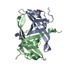 1v1qS S: Starting model for refinement |
|---|---|
| Similar structure data |


-Links
 PDBj
PDBj


- Assembly
| Deposited unit | 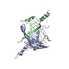
| ||||||||
|---|---|---|---|---|---|---|---|---|---|
| 1 |
| ||||||||
| Unit cell |
|
-Components
| #1: Protein | Mass: 13685.733 Da / Num. of mol.: 2 / Mutation: YES Source method: isolated from a genetically manipulated source Source: (gene. exp.) ESCHERICHIA COLI (E. coli) / Strain: K12 / Production host: ESCHERICHIA COLI (E. coli) / Strain (production host): BL21 / References: UniProt: P07013#2: DNA chain | | Mass: 4517.935 Da / Num. of mol.: 1 / Source method: obtained synthetically #3: Water | ChemComp-HOH / | Water Mass: 18.015 Da / Num. of mol.: 119 / Source method: isolated from a natural source / Formula: H2O Mass: 18.015 Da / Num. of mol.: 119 / Source method: isolated from a natural source / Formula: H2OCompound details | ENGINEERED | Sequence details | THE RECOMBINANT FORM OF PRIB CONTAINS PARTIAL T7 TAG IN N- TERMINAL END, AND HIS TAG IN C-TERMINAL ...THE RECOMBINAN | |
|---|
-Experimental details
-Experiment
| Experiment | Method: X-RAY DIFFRACTION / Number of used crystals: 1 |
|---|
- Sample preparation
| Crystal | Density Matthews: 1.8 Å3/Da / Density % sol: 31 % |
|---|---|
| Crystal grow | pH: 6.5 / Details: pH 6.50 |
-Data collection
| Diffraction | Mean temperature: 100 K |
|---|---|
| Diffraction source | Source: SYNCHROTRON / Site: NSRRC  / Beamline: BL17B2 / Wavelength: 1.12714 / Beamline: BL17B2 / Wavelength: 1.12714 |
| Detector | Type: MARRESEARCH / Detector: IMAGE PLATE / Date: Apr 30, 2005 / Details: MIRRORS |
| Radiation | Monochromator: DCM WITH SAGITTAL FOCUSING / Protocol: SINGLE WAVELENGTH / Monochromatic (M) / Laue (L): M / Scattering type: x-ray |
| Radiation wavelength | Wavelength: 1.12714 Å / Relative weight: 1 |
| Reflection | Resolution: 2.7→20 Å / Num. obs: 6773 / % possible obs: 99.6 % / Observed criterion σ(I): 2 / Redundancy: 4.9 % / Biso Wilson estimate: 44 Å2 / Rmerge(I) obs: 0.07 / Net I/σ(I): 30.34 |
| Reflection shell | Resolution: 2.7→2.8 Å / Redundancy: 4.7 % / Rmerge(I) obs: 0.42 / Mean I/σ(I) obs: 4.7 / % possible all: 100 |
- Processing
| Software |
| ||||||||||||||||||||||||||||||||||||||||||||||||||||||||||||||||||||||||||||||||
|---|---|---|---|---|---|---|---|---|---|---|---|---|---|---|---|---|---|---|---|---|---|---|---|---|---|---|---|---|---|---|---|---|---|---|---|---|---|---|---|---|---|---|---|---|---|---|---|---|---|---|---|---|---|---|---|---|---|---|---|---|---|---|---|---|---|---|---|---|---|---|---|---|---|---|---|---|---|---|---|---|---|
| Refinement | Method to determine structure: MOLECULAR REPLACEMENT Starting model: PDB ENTRY 1V1Q Resolution: 2.7→17.6 Å / Rfactor Rfree error: 0.011 / Data cutoff high absF: 270117.32 / Isotropic thermal model: RESTRAINED / Cross valid method: THROUGHOUT / σ(F): 2 / Stereochemistry target values: MAXIMUM LIKELIHOOD Details: 1. HIS-116 AND HIS-117 OF CHAIN A ARE INVISIBLE. 2. MET-6, ASP-5, HIS-115, HIS-116,HIS-117 OF CHAIN B ARE INVISIBLE. 3. THOSE SIDECHAINS LISTED AS FOLLOWS ARE DISORDERED CHAIN A MET-6, ASP- ...Details: 1. HIS-116 AND HIS-117 OF CHAIN A ARE INVISIBLE. 2. MET-6, ASP-5, HIS-115, HIS-116,HIS-117 OF CHAIN B ARE INVISIBLE. 3. THOSE SIDECHAINS LISTED AS FOLLOWS ARE DISORDERED CHAIN A MET-6, ASP-5, LEU-1, LEU-16, SER-20, GLN-45, ASN-59, HIS-81, MET-90, ILE-97, ILE-100, ASP-101, LEU- 106, HIS-112, HIS-114, HIS-115. CHAIN B SER-2, LEU-1, ASN-3, ILE-24, GLU-39, ILE-62, LEU-87, ASP-101, LYS-105, LEU-110.
| ||||||||||||||||||||||||||||||||||||||||||||||||||||||||||||||||||||||||||||||||
| Solvent computation | Solvent model: FLAT MODEL / Bsol: 59.3093 Å2 / ksol: 0.241866 e/Å3 | ||||||||||||||||||||||||||||||||||||||||||||||||||||||||||||||||||||||||||||||||
| Displacement parameters | Biso mean: 41 Å2
| ||||||||||||||||||||||||||||||||||||||||||||||||||||||||||||||||||||||||||||||||
| Refine analyze |
| ||||||||||||||||||||||||||||||||||||||||||||||||||||||||||||||||||||||||||||||||
| Refinement step | Cycle: LAST / Resolution: 2.7→17.6 Å
| ||||||||||||||||||||||||||||||||||||||||||||||||||||||||||||||||||||||||||||||||
| Refine LS restraints |
| ||||||||||||||||||||||||||||||||||||||||||||||||||||||||||||||||||||||||||||||||
| LS refinement shell | Resolution: 2.7→2.87 Å / Rfactor Rfree error: 0.035 / Total num. of bins used: 6
| ||||||||||||||||||||||||||||||||||||||||||||||||||||||||||||||||||||||||||||||||
| Xplor file |
|
