 Movie
Movie Controller

[English] 日本語
 Yorodumi
- PDB-1v1q: Crystal structure of PriB- a primosomal DNA replication protein o... -
+ Open data

- Basic information
| Entry | Database: PDB / ID: 1v1q | ||||||
|---|---|---|---|---|---|---|---|
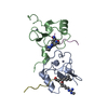
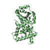
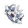
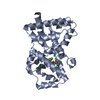
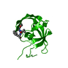
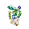
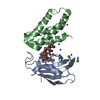
| Title | Crystal structure of PriB- a primosomal DNA replication protein of Escherichia coli | ||||||
 Components Components | PRIMOSOMAL REPLICATION PROTEIN N | ||||||
 Keywords Keywords |  DNA BINDING / PRIMOSOME / DNA REPLICATION DNA BINDING / PRIMOSOME / DNA REPLICATION | ||||||
| Function / homology |  Function and homology information Function and homology informationpre-primosome complex / DnaB-DnaC-DnaT-PriA-PriB complex / plasmid maintenance / primosome complex / DNA replication, synthesis of primer / replication fork processing / DNA unwinding involved in DNA replication / DNA replication initiation / response to radiation / single-stranded DNA binding / identical protein bindingSimilarity search - Function | ||||||
| Biological species |  ESCHERICHIA COLI (E. coli) ESCHERICHIA COLI (E. coli) | ||||||
| Method | X-RAY DIFFRACTION / SYNCHROTRON / MAD / Resolution: 2.1 Å | ||||||
 Authors Authors | Liu, J.-H. / Chang, T.-W. / Huang, C.-Y. / Chang, M.-C. / Chen, S.-U. / Wu, H.-N. / Hsiao, C.-D. | ||||||
 Citation Citation | Journal: J.Biol.Chem. / Year: 2004 Title: Crystal Structure of Prib- a Primosomal DNA Replication Protein of Escherichia Coli Authors: Liu, J.-H. / Chang, T.-W. / Huang, C.-Y. / Chen, S.-U. / Wu, H.-N. / Chang, M.-C. / Hsiao, C.-D. | ||||||
| History |
| ||||||
| Remark 700 | SHEET DETERMINATION METHOD: DSSP THE SHEETS PRESENTED AS "AA" IN EACH CHAIN ON SHEET RECORDS BELOW ... SHEET DETERMINATION METHOD: DSSP THE SHEETS PRESENTED AS "AA" IN EACH CHAIN ON SHEET RECORDS BELOW IS ACTUALLY AN 11-STRANDED BARREL THIS IS REPRESENTED BY A 12-STRANDED SHEET IN WHICH THE FIRST AND LAST STRANDS ARE IDENTICAL. |
- Structure visualization
| Structure viewer | Molecule: MolmilJmol/JSmol |
|---|
- Downloads & links
-Download
| PDBx/mmCIF format | 1v1q.cif.gz | 59.4 KB | Display | PDBx/mmCIF format |
|---|---|---|---|---|
| PDB format | pdb1v1q.ent.gz | 42.5 KB | Display | PDB format |
| PDBx/mmJSON format | 1v1q.json.gz | Tree view | PDBx/mmJSON format | |
| Others |  Other downloads Other downloads |
-Validation report
| Arichive directory | https://data.pdbj.org/pub/pdb/validation_reports/v1/1v1qftp://data.pdbj.org/pub/pdb/validation_reports/v1/1v1q | HTTPS FTP |
|---|
-Related structure data
| Similar structure data |
|---|


-Links
 PDBj
PDBj

- Assembly
| Deposited unit | 
| ||||||||
|---|---|---|---|---|---|---|---|---|---|
| 1 |
| ||||||||
| Unit cell |
|
-Components
| #1: Protein | Mass: 14791.990 Da / Num. of mol.: 2 / Mutation: YES Source method: isolated from a genetically manipulated source Source: (gene. exp.) ESCHERICHIA COLI (E. coli) / Strain: K12 / Plasmid: PET21B-PRIB / Production host: ESCHERICHIA COLI (E. coli) / Strain (production host): BL21(DE3) / References: UniProt: P07013#2: Chemical | Cysteine  Type: L-peptide linking / Mass: 121.158 Da / Num. of mol.: 3 / Source method: obtained synthetically / Formula: C3H7NO2S Type: L-peptide linking / Mass: 121.158 Da / Num. of mol.: 3 / Source method: obtained synthetically / Formula: C3H7NO2S#3: Water | ChemComp-HOH / | Water Mass: 18.015 Da / Num. of mol.: 134 / Source method: isolated from a natural source / Formula: H2O Mass: 18.015 Da / Num. of mol.: 134 / Source method: isolated from a natural source / Formula: H2OCompound details | ENGINEERED | Sequence details | RESIDUES PRECEDING POSITION 1 OF EACH MOLECULE ARE FROM T7-TAG, AND RESIDUES AFTER POSITION 104 OF ...RESIDUES PRECEDING POSITION 1 OF EACH MOLECULE ARE FROM T7-TAG, AND RESIDUES AFTER POSITION 104 OF CHAIN A ARE FROM HIS-TAG | |
|---|
-Experimental details
-Experiment
| Experiment | Method: X-RAY DIFFRACTION / Number of used crystals: 1 |
|---|
- Sample preparation
| Crystal | Density Matthews: 1.9 Å3/Da / Density % sol: 33.7 % |
|---|---|
| Crystal grow | pH: 5.7 / Details: pH 5.70 |
-Data collection
| Diffraction | Mean temperature: 110 K | |||||||||||||||
|---|---|---|---|---|---|---|---|---|---|---|---|---|---|---|---|---|
| Diffraction source | Source: SYNCHROTRON / Site: NSRRC  / Beamline: BL17B2 / Wavelength: 1.0083, 1.0093, 0.9921, 1.0249 / Beamline: BL17B2 / Wavelength: 1.0083, 1.0093, 0.9921, 1.0249 | |||||||||||||||
| Detector | Type: RIGAKU IMAGE PLATE / Detector: IMAGE PLATE / Details: MIRRORS | |||||||||||||||
| Radiation | Protocol: MAD / Monochromatic (M) / Laue (L): M / Scattering type: x-ray | |||||||||||||||
| Radiation wavelength |
| |||||||||||||||
| Reflection | Resolution: 2.1→23.17 Å / Num. obs: 13751 / % possible obs: 99.9 % / Observed criterion σ(I): 2 / Redundancy: 3.6 % / Biso Wilson estimate: 13.9 Å2 / Rmerge(I) obs: 0.083 / Net I/σ(I): 14.1 | |||||||||||||||
| Reflection shell | Resolution: 2.1→2.2 Å / Rmerge(I) obs: 0.561 / Mean I/σ(I) obs: 2.2 / % possible all: 99.6 |
- Processing
| Software |
| ||||||||||||||||||||||||||||||||||||||||||||||||||||||||||||||||||||||||||||||||
|---|---|---|---|---|---|---|---|---|---|---|---|---|---|---|---|---|---|---|---|---|---|---|---|---|---|---|---|---|---|---|---|---|---|---|---|---|---|---|---|---|---|---|---|---|---|---|---|---|---|---|---|---|---|---|---|---|---|---|---|---|---|---|---|---|---|---|---|---|---|---|---|---|---|---|---|---|---|---|---|---|---|
| Refinement | Method to determine structure: MAD / Resolution: 2.1→23.17 Å / Rfactor Rfree error: 0.008 / Data cutoff high absF: 284509.74 / Isotropic thermal model: RESTRAINED / Cross valid method: THROUGHOUT / σ(F): 2
| ||||||||||||||||||||||||||||||||||||||||||||||||||||||||||||||||||||||||||||||||
| Solvent computation | Solvent model: FLAT / Bsol: 40.8918 Å2 / ksol: 0.312944 e/Å3 | ||||||||||||||||||||||||||||||||||||||||||||||||||||||||||||||||||||||||||||||||
| Displacement parameters | Biso mean: 40.1 Å2
| ||||||||||||||||||||||||||||||||||||||||||||||||||||||||||||||||||||||||||||||||
| Refine analyze |
| ||||||||||||||||||||||||||||||||||||||||||||||||||||||||||||||||||||||||||||||||
| Refinement step | Cycle: LAST / Resolution: 2.1→23.17 Å
| ||||||||||||||||||||||||||||||||||||||||||||||||||||||||||||||||||||||||||||||||
| Refine LS restraints |
| ||||||||||||||||||||||||||||||||||||||||||||||||||||||||||||||||||||||||||||||||
| LS refinement shell | Resolution: 2.1→2.23 Å / Rfactor Rfree error: 0.026 / Total num. of bins used: 6
| ||||||||||||||||||||||||||||||||||||||||||||||||||||||||||||||||||||||||||||||||
| Xplor file |
|
