 Movie
Movie Controller

[English] 日本語
 Yorodumi
- PDB-3kty: Crystal Structure of Probable Methyltransferase from Bordetella p... -
+ Open data

- Basic information
| Entry | Database: PDB / ID: 3kty | ||||||
|---|---|---|---|---|---|---|---|
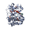
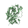
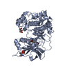
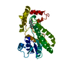
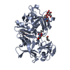
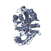
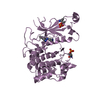
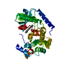
| Title | Crystal Structure of Probable Methyltransferase from Bordetella pertussis Tohama I | ||||||
 Components Components | Probable methyltransferase | ||||||
 Keywords Keywords |  TRANSFERASE / alpha-beta-alpha sandwich / Structural Genomics / PSI-2 / Protein Structure Initiative / Midwest Center for Structural Genomics / MCSG / Methyltransferase TRANSFERASE / alpha-beta-alpha sandwich / Structural Genomics / PSI-2 / Protein Structure Initiative / Midwest Center for Structural Genomics / MCSG / Methyltransferase | ||||||
| Function / homology |  Function and homology information Function and homology informationRNA methyltransferase activity / RNA processing / RNA binding / cytoplasmSimilarity search - Function | ||||||
| Biological species |  Bordetella pertussis (bacteria) Bordetella pertussis (bacteria) | ||||||
| Method | X-RAY DIFFRACTION / SYNCHROTRON / SAD / Resolution: 2.302 Å | ||||||
 Authors Authors | Kim, Y. / Tesar, C. / Keigher, L. / Joachimiak, A. / Midwest Center for Structural Genomics (MCSG) | ||||||
 Citation Citation | Journal: To be Published Title: Crystal Structure of Probable Methyltransferase SpoU from Bordetella pertussis Tohama I Authors: Kim, Y. / Tesar, C. / Keigher, L. / Joachimiak, A. | ||||||
| History |
|
- Structure visualization
| Structure viewer | Molecule: MolmilJmol/JSmol |
|---|
- Downloads & links
-Download
| PDBx/mmCIF format | 3kty.cif.gz | 209.1 KB | Display | PDBx/mmCIF format |
|---|---|---|---|---|
| PDB format | pdb3kty.ent.gz | 176.9 KB | Display | PDB format |
| PDBx/mmJSON format | 3kty.json.gz | Tree view | PDBx/mmJSON format | |
| Others |  Other downloads Other downloads |
-Validation report
| Arichive directory | https://data.pdbj.org/pub/pdb/validation_reports/kt/3ktyftp://data.pdbj.org/pub/pdb/validation_reports/kt/3kty | HTTPS FTP |
|---|
-Related structure data
| Similar structure data | |
|---|---|
| Other databases |


-Links
 PDBj
PDBj- Assembly
| Deposited unit | 
| ||||||||
|---|---|---|---|---|---|---|---|---|---|
| 1 | 
| ||||||||
| 2 | 
| ||||||||
| Unit cell |
|
-Components
| #1: Protein | Mass: 18665.768 Da / Num. of mol.: 3 / Fragment: target domain 1 - 170 Source method: isolated from a genetically manipulated source Source: (gene. exp.) Bordetella pertussis (bacteria) / Strain: Tohama I / Gene: BP1901 / Plasmid: pMCSG19 / Production host: Escherichia coli (E. coli) / Strain (production host): BL21 Magic / References: UniProt: Q7VXA3#2: Chemical | Glycerol  Mass: 92.094 Da / Num. of mol.: 2 / Source method: obtained synthetically / Formula: C3H8O3 Mass: 92.094 Da / Num. of mol.: 2 / Source method: obtained synthetically / Formula: C3H8O3#3: Chemical | Sulfate  Mass: 96.063 Da / Num. of mol.: 2 / Source method: obtained synthetically / Formula: SO4 Mass: 96.063 Da / Num. of mol.: 2 / Source method: obtained synthetically / Formula: SO4#4: Water | ChemComp-HOH / | Water Mass: 18.015 Da / Num. of mol.: 128 / Source method: isolated from a natural source / Formula: H2O Mass: 18.015 Da / Num. of mol.: 128 / Source method: isolated from a natural source / Formula: H2O |
|---|
-Experimental details
-Experiment
| Experiment | Method: X-RAY DIFFRACTION / Number of used crystals: 1 |
|---|
- Sample preparation
| Crystal | Density Matthews: 2.3 Å3/Da / Density % sol: 46.41 % |
|---|---|
| Crystal grow | Temperature: 297 K / Method: vapor diffusion, sitting drop / pH: 7.5 Details: 0.2 M Lithium sulfate monohydrate, 0.1 M HEPES pH7.5, 25 % w/v Polyehtlyene glycol 3,350, VAPOR DIFFUSION, SITTING DROP, temperature 297K |
-Data collection
| Diffraction | Mean temperature: 100 K |
|---|---|
| Diffraction source | Source: SYNCHROTRON / Site: APS  / Beamline: 19-ID / Wavelength: 0.9794 Å / Beamline: 19-ID / Wavelength: 0.9794 Å |
| Detector | Type: ADSC QUANTUM 315r / Detector: CCD / Date: Nov 14, 2008 / Details: mirrors |
| Radiation | Monochromator: double crystal monochromator / Protocol: SINGLE WAVELENGTH / Monochromatic (M) / Laue (L): M / Scattering type: x-ray |
| Radiation wavelength | Wavelength: 0.9794 Å / Relative weight: 1 |
| Reflection | Resolution: 2.3→43.52 Å / Num. all: 22535 / Num. obs: 22535 / % possible obs: 99.6 % / Observed criterion σ(F): 0 / Observed criterion σ(I): 0 / Redundancy: 4.6 % / Biso Wilson estimate: 43.52 Å2 / Rsym value: 0.117 / Net I/σ(I): 8.7 |
| Reflection shell | Resolution: 2.3→2.34 Å / Redundancy: 3.5 % / Mean I/σ(I) obs: 1.7 / Num. unique all: 1130 / Rsym value: 0.623 / % possible all: 97.5 |
- Processing
| Software |
| ||||||||||||||||||||||||||||||||||||||||||||||||||||||||||||||||||||||||||||||||||||||||||||||||||||||||
|---|---|---|---|---|---|---|---|---|---|---|---|---|---|---|---|---|---|---|---|---|---|---|---|---|---|---|---|---|---|---|---|---|---|---|---|---|---|---|---|---|---|---|---|---|---|---|---|---|---|---|---|---|---|---|---|---|---|---|---|---|---|---|---|---|---|---|---|---|---|---|---|---|---|---|---|---|---|---|---|---|---|---|---|---|---|---|---|---|---|---|---|---|---|---|---|---|---|---|---|---|---|---|---|---|---|
| Refinement | Method to determine structure: SAD / Resolution: 2.302→43.52 Å / SU ML: 0.36 / Isotropic thermal model: mixed / Cross valid method: THROUGHOUT / σ(F): 0 / Phase error: 27.19 / Stereochemistry target values: MLHL
| ||||||||||||||||||||||||||||||||||||||||||||||||||||||||||||||||||||||||||||||||||||||||||||||||||||||||
| Solvent computation | Shrinkage radii: 0.9 Å / VDW probe radii: 1.11 Å / Solvent model: FLAT BULK SOLVENT MODEL / Bsol: 45.368 Å2 / ksol: 0.316 e/Å3 | ||||||||||||||||||||||||||||||||||||||||||||||||||||||||||||||||||||||||||||||||||||||||||||||||||||||||
| Displacement parameters |
| ||||||||||||||||||||||||||||||||||||||||||||||||||||||||||||||||||||||||||||||||||||||||||||||||||||||||
| Refinement step | Cycle: LAST / Resolution: 2.302→43.52 Å
| ||||||||||||||||||||||||||||||||||||||||||||||||||||||||||||||||||||||||||||||||||||||||||||||||||||||||
| Refine LS restraints |
| ||||||||||||||||||||||||||||||||||||||||||||||||||||||||||||||||||||||||||||||||||||||||||||||||||||||||
| LS refinement shell | Refine-ID: X-RAY DIFFRACTION
| ||||||||||||||||||||||||||||||||||||||||||||||||||||||||||||||||||||||||||||||||||||||||||||||||||||||||
| Refinement TLS params. | Refine-ID: X-RAY DIFFRACTION
| ||||||||||||||||||||||||||||||||||||||||||||||||||||||||||||||||||||||||||||||||||||||||||||||||||||||||
| Refinement TLS group | Selection details: chain C |
