 Movie
Movie Controller


+ Open data

- Basic information
| Entry | Database: PDB / ID: 3k8a | ||||||
|---|---|---|---|---|---|---|---|
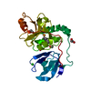
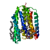
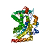
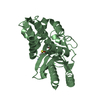
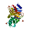
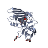
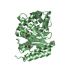
| Title | Neisseria gonorrhoeae PriB | ||||||
 Components Components | Putative primosomal replication protein | ||||||
 Keywords Keywords |  DNA BINDING PROTEIN / beta-barrel / OB-fold DNA BINDING PROTEIN / beta-barrel / OB-fold | ||||||
| Function / homology |  Function and homology information Function and homology informationpre-primosome complex / positive regulation of helicase activity / DNA replication, synthesis of primer / positive regulation of ATP-dependent activity / ATPase activator activity / single-stranded DNA binding / ATPase binding / double-stranded DNA binding / protein homodimerization activitySimilarity search - Function | ||||||
| Biological species |  Neisseria gonorrhoeae FA 1090 (bacteria) Neisseria gonorrhoeae FA 1090 (bacteria) | ||||||
| Method | X-RAY DIFFRACTION / SYNCHROTRON / MOLECULAR REPLACEMENT / molecular replacement / Resolution: 2.7 Å | ||||||
 Authors Authors | Lopper, M.E. / Dong, J. / George, N.P. / Duckett, K.L. / DeBeer, M.A. | ||||||
 Citation Citation | Journal: Nucleic Acids Res. / Year: 2010 Title: The crystal structure of Neisseria gonorrhoeae PriB reveals mechanistic differences among bacterial DNA replication restart pathways Authors: Dong, J. / George, N.P. / Duckett, K.L. / DeBeer, M.A. / Lopper, M.E. | ||||||
| History |
|
- Structure visualization
| Structure viewer | Molecule: MolmilJmol/JSmol |
|---|
- Downloads & links
-Download
| PDBx/mmCIF format | 3k8a.cif.gz | 52.9 KB | Display | PDBx/mmCIF format |
|---|---|---|---|---|
| PDB format | pdb3k8a.ent.gz | 38 KB | Display | PDB format |
| PDBx/mmJSON format | 3k8a.json.gz | Tree view | PDBx/mmJSON format | |
| Others |  Other downloads Other downloads |
-Validation report
| Arichive directory | https://data.pdbj.org/pub/pdb/validation_reports/k8/3k8aftp://data.pdbj.org/pub/pdb/validation_reports/k8/3k8a | HTTPS FTP |
|---|
-Related structure data
| Related structure data | 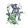 1v1qS S: Starting model for refinement |
|---|---|
| Similar structure data |


-Links
 PDBj
PDBj- Assembly
| Deposited unit | 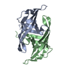
| ||||||||
|---|---|---|---|---|---|---|---|---|---|
| 1 |
| ||||||||
| Unit cell |
|
-Components
| #1: Protein | Mass: 11740.555 Da / Num. of mol.: 2 Source method: isolated from a genetically manipulated source Source: (gene. exp.) Neisseria gonorrhoeae FA 1090 (bacteria)Strain: FA1090 / Gene: NGO0582, prib / Plasmid: pET28b / Production host: Escherichia coli (E. coli) / Strain (production host): BL21(DE3) / References: UniProt: Q5F924#2: Water | ChemComp-HOH / | Water Mass: 18.015 Da / Num. of mol.: 8 / Source method: isolated from a natural source / Formula: H2O Mass: 18.015 Da / Num. of mol.: 8 / Source method: isolated from a natural source / Formula: H2O |
|---|
-Experimental details
-Experiment
| Experiment | Method: X-RAY DIFFRACTION / Number of used crystals: 1 |
|---|
- Sample preparation
| Crystal | Density Matthews: 4.12 Å3/Da / Density % sol: 70.13 % |
|---|---|
| Crystal grow | Temperature: 298 K / Method: vapor diffusion, hanging drop / pH: 4.2 Details: 4% (w/v) PEG 4000, pH 4.2, VAPOR DIFFUSION, HANGING DROP, temperature 298K |
-Data collection
| Diffraction | Mean temperature: 110 K |
|---|---|
| Diffraction source | Source: SYNCHROTRON / Site: APS  / Beamline: 21-ID-F / Wavelength: 0.97872 Å / Beamline: 21-ID-F / Wavelength: 0.97872 Å |
| Detector | Type: MARMOSAIC 225 mm CCD / Detector: CCD / Date: Dec 11, 2008 |
| Radiation | Monochromator: SI(111) DOUBLE CRYSTAL / Protocol: SINGLE WAVELENGTH / Monochromatic (M) / Laue (L): M / Scattering type: x-ray |
| Radiation wavelength | Wavelength: 0.97872 Å / Relative weight: 1 |
| Reflection | Resolution: 2.7→30 Å / Num. all: 10124 / Num. obs: 9741 / % possible obs: 92.8 % / Observed criterion σ(F): 0 / Observed criterion σ(I): 0 / Redundancy: 13.3 % / Biso Wilson estimate: 68.2 Å2 / Rmerge(I) obs: 0.072 / Rsym value: 0.072 / Χ2: 1.058 / Net I/σ(I): 27 |
| Reflection shell | Resolution: 2.7→2.75 Å / Redundancy: 10.4 % / Rmerge(I) obs: 0.22 / Mean I/σ(I) obs: 5.5 / Num. unique all: 354 / Rsym value: 0.22 / Χ2: 1.136 / % possible all: 65.2 |
-Phasing
| Phasing | Method: molecular replacement | |||||||||
|---|---|---|---|---|---|---|---|---|---|---|
| Phasing MR | Rfactor: 33.07 / Model details: Phaser MODE: MR_AUTO
|
- Processing
| Software |
| |||||||||||||||||||||||||||||||||||||||||||||||||||||||||||||||||||||||||||
|---|---|---|---|---|---|---|---|---|---|---|---|---|---|---|---|---|---|---|---|---|---|---|---|---|---|---|---|---|---|---|---|---|---|---|---|---|---|---|---|---|---|---|---|---|---|---|---|---|---|---|---|---|---|---|---|---|---|---|---|---|---|---|---|---|---|---|---|---|---|---|---|---|---|---|---|---|
| Refinement | Method to determine structure: MOLECULAR REPLACEMENT Starting model: PDB entry 1V1Q Resolution: 2.7→26.25 Å / Cor.coef. Fo:Fc: 0.934 / Cor.coef. Fo:Fc free: 0.917 / WRfactor Rfree: 0.357 / WRfactor Rwork: 0.294 / Occupancy max: 1 / Occupancy min: 1 / FOM work R set: 0.855 / SU B: 32.3 / SU ML: 0.293 / SU R Cruickshank DPI: 0.534 / SU Rfree: 0.342 / TLS residual ADP flag: LIKELY RESIDUAL / Isotropic thermal model: ISOTROPIC / Cross valid method: THROUGHOUT / σ(F): 0 / ESU R: 0.534 / ESU R Free: 0.341 Stereochemistry target values: MAXIMUM LIKELIHOOD WITH PHASES Details: HYDROGENS HAVE BEEN ADDED IN THE RIDING POSITIONS U VALUES: RESIDUAL ONLY
| |||||||||||||||||||||||||||||||||||||||||||||||||||||||||||||||||||||||||||
| Solvent computation | Ion probe radii: 0.8 Å / Shrinkage radii: 0.8 Å / VDW probe radii: 1.4 Å / Solvent model: MASK | |||||||||||||||||||||||||||||||||||||||||||||||||||||||||||||||||||||||||||
| Displacement parameters | Biso max: 91.7 Å2 / Biso mean: 56.509 Å2 / Biso min: 13.64 Å2
| |||||||||||||||||||||||||||||||||||||||||||||||||||||||||||||||||||||||||||
| Refinement step | Cycle: LAST / Resolution: 2.7→26.25 Å
| |||||||||||||||||||||||||||||||||||||||||||||||||||||||||||||||||||||||||||
| Refine LS restraints |
| |||||||||||||||||||||||||||||||||||||||||||||||||||||||||||||||||||||||||||
| LS refinement shell | Resolution: 2.7→2.769 Å / Total num. of bins used: 20
| |||||||||||||||||||||||||||||||||||||||||||||||||||||||||||||||||||||||||||
| Refinement TLS params. | Method: refined / Refine-ID: X-RAY DIFFRACTION
| |||||||||||||||||||||||||||||||||||||||||||||||||||||||||||||||||||||||||||
| Refinement TLS group |
|
