 Movie
Movie Controller


+ Open data

- Basic information
| Entry | Database: PDB / ID: 3k6f | ||||||
|---|---|---|---|---|---|---|---|
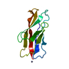
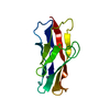
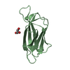
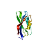
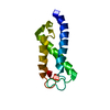
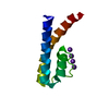
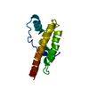
| Title | Crystal structure of mouse T-cadherin EC1 | ||||||
 Components Components | T-cadherin | ||||||
 Keywords Keywords | CELL ADHESION / T-cadherin / Calcium / Cell membrane / Cleavage on pair of basic residues / Glycoprotein / GPI-anchor / Lipoprotein / Membrane | ||||||
| Function / homology |  Function and homology information Function and homology informationlow-density lipoprotein particle mediated signaling / adiponectin binding / regulation of epidermal growth factor receptor signaling pathway / Adherens junctions interactions / localization within membrane / cell-cell adhesion mediated by cadherin / low-density lipoprotein particle binding / calcium-dependent cell-cell adhesion via plasma membrane cell adhesion molecules / negative regulation of cell adhesion / catenin complex ...low-density lipoprotein particle mediated signaling / adiponectin binding / regulation of epidermal growth factor receptor signaling pathway / Adherens junctions interactions / localization within membrane / cell-cell adhesion mediated by cadherin / low-density lipoprotein particle binding / calcium-dependent cell-cell adhesion via plasma membrane cell adhesion molecules / negative regulation of cell adhesion / catenin complex / cell-cell junction assembly / adherens junction organization / sprouting angiogenesis / positive regulation of positive chemotaxis / lamellipodium assembly / positive regulation of cell-matrix adhesion / Rac protein signal transduction / homophilic cell adhesion via plasma membrane adhesion molecules / lipoprotein particle binding / Rho protein signal transduction / regulation of endocytosis / keratinocyte proliferation / endothelial cell migration / GABA-ergic synapse / positive regulation of calcium-mediated signaling / positive regulation of endothelial cell proliferation / caveola / adherens junction / positive regulation of smooth muscle cell proliferation / cell morphogenesis / neuron projection / positive regulation of cell migration / cadherin binding / negative regulation of cell population proliferation / external side of plasma membrane / synapse / calcium ion binding / perinuclear region of cytoplasm / positive regulation of transcription by RNA polymerase II / extracellular space / identical protein binding / plasma membrane / cytoplasmSimilarity search - Function | ||||||
| Biological species |  Mus musculus (house mouse) Mus musculus (house mouse) | ||||||
| Method | X-RAY DIFFRACTION / SYNCHROTRON / MOLECULAR REPLACEMENT / Resolution: 1.813 Å | ||||||
 Authors Authors | Shapiro, L. / Ciatto, C. | ||||||
 Citation Citation | Journal: Nat.Struct.Mol.Biol. / Year: 2010 Title: T-cadherin structures reveal a novel adhesive binding mechanism Authors: Ciatto, C. / Bahna, F. / Zampieri, N. / Vansteenhouse, H.C. / Katsamba, P.S. / Ahlsen, G. / Harrison, O.J. / Brasch, J. / Jin, X. / Posy, S. / Vendome, J. / Ranscht, B. / Jessell, T.M. / Honig, B. / Shapiro, L. | ||||||
| History |
|
- Structure visualization
| Structure viewer | Molecule: MolmilJmol/JSmol |
|---|
- Downloads & links
-Download
| PDBx/mmCIF format | 3k6f.cif.gz | 91.6 KB | Display | PDBx/mmCIF format |
|---|---|---|---|---|
| PDB format | pdb3k6f.ent.gz | 71.2 KB | Display | PDB format |
| PDBx/mmJSON format | 3k6f.json.gz | Tree view | PDBx/mmJSON format | |
| Others |  Other downloads Other downloads |
-Validation report
| Arichive directory | https://data.pdbj.org/pub/pdb/validation_reports/k6/3k6fftp://data.pdbj.org/pub/pdb/validation_reports/k6/3k6f | HTTPS FTP |
|---|
-Related structure data
| Related structure data | 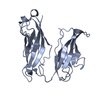 3k5rC  3k5sC  3k6dSC  3k6iC S: Starting model for refinement C: citing same article ( |
|---|---|
| Similar structure data |


-Links
 PDBj
PDBj
- Assembly
| Deposited unit | 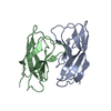
| |||||||||||||||||||||
|---|---|---|---|---|---|---|---|---|---|---|---|---|---|---|---|---|---|---|---|---|---|---|
| 1 | 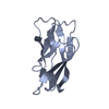
| |||||||||||||||||||||
| 2 | 
| |||||||||||||||||||||
| Unit cell |
| |||||||||||||||||||||
| Components on special symmetry positions |
|
-Components
| #1: Protein | / Truncated cadherin / Cadherin-13 / T-cad / Heart cadherin / H-cadherin Mass: 10925.205 Da / Num. of mol.: 2 / Fragment: EC1 domain: UNP residues 140-237 Source method: isolated from a genetically manipulated source Source: (gene. exp.) Mus musculus (house mouse) / Gene: Cdh13 / Production host:  Escherichia coli (E. coli) / References: UniProt: Q9WTR5 Escherichia coli (E. coli) / References: UniProt: Q9WTR5#2: Water | ChemComp-HOH / | Water Mass: 18.015 Da / Num. of mol.: 203 / Source method: isolated from a natural source / Formula: H2O Mass: 18.015 Da / Num. of mol.: 203 / Source method: isolated from a natural source / Formula: H2O |
|---|
-Experimental details
-Experiment
| Experiment | Method: X-RAY DIFFRACTION / Number of used crystals: 1 |
|---|
- Sample preparation
| Crystal | Density Matthews: 2.05 Å3/Da / Density % sol: 40.03 % |
|---|---|
| Crystal grow | Temperature: 277 K / Method: vapor diffusion, hanging drop / pH: 6 Details: 0.2M Ammonium sulfate, 20% PEG 3350, pH 6.0, VAPOR DIFFUSION, HANGING DROP, temperature 277K |
-Data collection
| Diffraction | Mean temperature: 100 K |
|---|---|
| Diffraction source | Source: SYNCHROTRON / Site: NSLS  / Beamline: X4C / Wavelength: 0.9791 Å / Beamline: X4C / Wavelength: 0.9791 Å |
| Detector | Type: MAR scanner 345 mm plate / Detector: IMAGE PLATE / Details: Vertical focusing mirror |
| Radiation | Monochromator: Bent single Si(111) crystal (horizontal focusing and deflection) Protocol: SINGLE WAVELENGTH / Monochromatic (M) / Laue (L): M / Scattering type: x-ray |
| Radiation wavelength | Wavelength: 0.9791 Å / Relative weight: 1 |
| Reflection | Resolution: 1.8→30 Å / Num. obs: 16137 / % possible obs: 99.3 % / Redundancy: 10.8 % / Biso Wilson estimate: 17.473 Å2 / Rmerge(I) obs: 0.048 |
| Reflection shell | Resolution: 1.8→1.9 Å / Rmerge(I) obs: 0.125 / % possible all: 99.9 |
- Processing
| Software |
| ||||||||||||||||||||||||||||||||||||||||||||||||||||||||||||||||||||||||||||||||||||||||||||||||||||||||||||||||||||||||||||||||||||||||||||||||||||||||||||||||||||||||||||||
|---|---|---|---|---|---|---|---|---|---|---|---|---|---|---|---|---|---|---|---|---|---|---|---|---|---|---|---|---|---|---|---|---|---|---|---|---|---|---|---|---|---|---|---|---|---|---|---|---|---|---|---|---|---|---|---|---|---|---|---|---|---|---|---|---|---|---|---|---|---|---|---|---|---|---|---|---|---|---|---|---|---|---|---|---|---|---|---|---|---|---|---|---|---|---|---|---|---|---|---|---|---|---|---|---|---|---|---|---|---|---|---|---|---|---|---|---|---|---|---|---|---|---|---|---|---|---|---|---|---|---|---|---|---|---|---|---|---|---|---|---|---|---|---|---|---|---|---|---|---|---|---|---|---|---|---|---|---|---|---|---|---|---|---|---|---|---|---|---|---|---|---|---|---|---|---|
| Refinement | Method to determine structure: MOLECULAR REPLACEMENT Starting model: PRB entry 3K6D Resolution: 1.813→19.447 Å / Occupancy max: 1 / Occupancy min: 1 / FOM work R set: 0.859 / Stereochemistry target values: MAXIMUM LIKELIHOOD
| ||||||||||||||||||||||||||||||||||||||||||||||||||||||||||||||||||||||||||||||||||||||||||||||||||||||||||||||||||||||||||||||||||||||||||||||||||||||||||||||||||||||||||||||
| Solvent computation | Bsol: 63.705 Å2 / ksol: 0.423 e/Å3 | ||||||||||||||||||||||||||||||||||||||||||||||||||||||||||||||||||||||||||||||||||||||||||||||||||||||||||||||||||||||||||||||||||||||||||||||||||||||||||||||||||||||||||||||
| Displacement parameters | Biso max: 77.38 Å2 / Biso mean: 22.144 Å2 / Biso min: 8.8 Å2
| ||||||||||||||||||||||||||||||||||||||||||||||||||||||||||||||||||||||||||||||||||||||||||||||||||||||||||||||||||||||||||||||||||||||||||||||||||||||||||||||||||||||||||||||
| Refinement step | Cycle: LAST / Resolution: 1.813→19.447 Å
| ||||||||||||||||||||||||||||||||||||||||||||||||||||||||||||||||||||||||||||||||||||||||||||||||||||||||||||||||||||||||||||||||||||||||||||||||||||||||||||||||||||||||||||||
| Refine LS restraints |
| ||||||||||||||||||||||||||||||||||||||||||||||||||||||||||||||||||||||||||||||||||||||||||||||||||||||||||||||||||||||||||||||||||||||||||||||||||||||||||||||||||||||||||||||
| LS refinement shell |
| ||||||||||||||||||||||||||||||||||||||||||||||||||||||||||||||||||||||||||||||||||||||||||||||||||||||||||||||||||||||||||||||||||||||||||||||||||||||||||||||||||||||||||||||
| Refinement TLS params. | Method: refined / Refine-ID: X-RAY DIFFRACTION
| ||||||||||||||||||||||||||||||||||||||||||||||||||||||||||||||||||||||||||||||||||||||||||||||||||||||||||||||||||||||||||||||||||||||||||||||||||||||||||||||||||||||||||||||
| Refinement TLS group |
|
