 Movie
Movie Controller


+ Open data

- Basic information
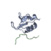
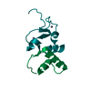
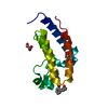
| Entry | Database: PDB / ID: 3k3v | ||||||
|---|---|---|---|---|---|---|---|
| Title | Crystal structure the GYF domain of S. Cerevisiae SMY2 | ||||||
 Components Components | Protein SMY2 | ||||||
 Keywords Keywords |  PROTEIN BINDING / GYF domain / poly-proline binding / domain swap / RAGNYA / SMY2 / Phosphoprotein PROTEIN BINDING / GYF domain / poly-proline binding / domain swap / RAGNYA / SMY2 / Phosphoprotein | ||||||
| Function / homology |  Function and homology information Function and homology informationendoplasmic reticulum to Golgi vesicle-mediated transport / endoplasmic reticulum membrane / cytosol / cytoplasmSimilarity search - Function | ||||||
| Biological species |  Saccharomyces cerevisiae (brewer's yeast) Saccharomyces cerevisiae (brewer's yeast) | ||||||
| Method | X-RAY DIFFRACTION / SYNCHROTRON / MOLECULAR REPLACEMENT / Resolution: 1.8 Å | ||||||
 Authors Authors | Ash, M.R. / Faelber, K. | ||||||
 Citation Citation | Journal: To be Published Title: SMY2-type GYF domain recognition in mRNA surveillance complexes Authors: Ash, M.R. / Faelber, K. / Kosslick, D. / Albert, G. / Roske, Y. / Freund, C. | ||||||
| History |
|
- Structure visualization
| Structure viewer | Molecule: MolmilJmol/JSmol |
|---|
- Downloads & links
-Download
| PDBx/mmCIF format | 3k3v.cif.gz | 31.6 KB | Display | PDBx/mmCIF format |
|---|---|---|---|---|
| PDB format | pdb3k3v.ent.gz | 20.4 KB | Display | PDB format |
| PDBx/mmJSON format | 3k3v.json.gz | Tree view | PDBx/mmJSON format | |
| Others |  Other downloads Other downloads |
-Validation report
| Arichive directory | https://data.pdbj.org/pub/pdb/validation_reports/k3/3k3vftp://data.pdbj.org/pub/pdb/validation_reports/k3/3k3v | HTTPS FTP |
|---|
-Related structure data
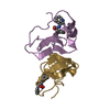
| Related structure data |  3fmaSC S: Starting model for refinement C: citing same article ( |
|---|---|
| Similar structure data |


-Links
 PDBj
PDBj- Assembly
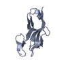
| Deposited unit | 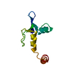
| ||||||||
|---|---|---|---|---|---|---|---|---|---|
| 1 |
| ||||||||
| 2 | 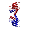
| ||||||||
| Unit cell |
|
-Components
| #1: Protein | Mass: 11109.328 Da / Num. of mol.: 1 / Fragment: GYF domain Source method: isolated from a genetically manipulated source Source: (gene. exp.) Saccharomyces cerevisiae (brewer's yeast)Gene: SMY2 / Plasmid: pET28 / Production host:  Escherichia coli BL21(DE3) (bacteria) / Strain (production host): BL21(DE3) / References: UniProt: P32909 Escherichia coli BL21(DE3) (bacteria) / Strain (production host): BL21(DE3) / References: UniProt: P32909 |
|---|---|
| #2: Water | ChemComp-HOH / Water Mass: 18.015 Da / Num. of mol.: 84 / Source method: isolated from a natural source / Formula: H2O Mass: 18.015 Da / Num. of mol.: 84 / Source method: isolated from a natural source / Formula: H2O |
-Experimental details
-Experiment
| Experiment | Method: X-RAY DIFFRACTION / Number of used crystals: 1 |
|---|
- Sample preparation
| Crystal | Density Matthews: 3.38 Å3/Da / Density % sol: 63.65 % |
|---|---|
| Crystal grow | Temperature: 293 K / Method: vapor diffusion, sitting drop / pH: 7.5 Details: 18% polyacrylic acid, 0.15M MgCl2, 0.1M Na-HEPES, pH 7.5, Vapor diffusion, sitting drop, temperature 293K |
-Data collection
| Diffraction | Mean temperature: 100 K |
|---|---|
| Diffraction source | Source: SYNCHROTRON / Site: BESSY  / Beamline: 14.2 / Wavelength: 0.91841 Å / Beamline: 14.2 / Wavelength: 0.91841 Å |
| Detector | Type: MAR CCD 165 mm / Detector: CCD / Date: Jul 19, 2006 |
| Radiation | Protocol: SINGLE WAVELENGTH / Monochromatic (M) / Laue (L): M / Scattering type: x-ray |
| Radiation wavelength | Wavelength: 0.91841 Å / Relative weight: 1 |
| Reflection | Resolution: 1.8→25.19 Å / Num. all: 14884 / Num. obs: 14884 / % possible obs: 99.8 % / Redundancy: 20.9 % / Biso Wilson estimate: 25.5 Å2 / Rmerge(I) obs: 0.075 / Net I/σ(I): 31.4 |
| Reflection shell | Resolution: 1.8→1.9 Å / Redundancy: 20.2 % / Rmerge(I) obs: 0.656 / Mean I/σ(I) obs: 5.2 / Num. measured all: 42500 / Num. unique all: 2107 / % possible all: 99.7 |
- Processing
| Software |
| |||||||||||||||||||||||||||||||||||||||||||||||||||||||||||||||||||||||||||
|---|---|---|---|---|---|---|---|---|---|---|---|---|---|---|---|---|---|---|---|---|---|---|---|---|---|---|---|---|---|---|---|---|---|---|---|---|---|---|---|---|---|---|---|---|---|---|---|---|---|---|---|---|---|---|---|---|---|---|---|---|---|---|---|---|---|---|---|---|---|---|---|---|---|---|---|---|
| Refinement | Method to determine structure: MOLECULAR REPLACEMENT Starting model: 3FMA Resolution: 1.8→25.19 Å / Cor.coef. Fo:Fc: 0.949 / Cor.coef. Fo:Fc free: 0.938 / Occupancy max: 1 / Occupancy min: 0.4 / SU B: 4.467 / SU ML: 0.063 / TLS residual ADP flag: LIKELY RESIDUAL / Isotropic thermal model: Isotropic with TLS / Cross valid method: THROUGHOUT / σ(F): 0 / ESU R: 0.099 / ESU R Free: 0.094 / Stereochemistry target values: MAXIMUM LIKELIHOOD Details: HYDROGENS HAVE BEEN ADDED IN THE RIDING POSITIONS U VALUES: RESIDUAL ONLY
| |||||||||||||||||||||||||||||||||||||||||||||||||||||||||||||||||||||||||||
| Solvent computation | Ion probe radii: 0.8 Å / Shrinkage radii: 0.8 Å / VDW probe radii: 1.2 Å / Solvent model: BABINET MODEL WITH MASK | |||||||||||||||||||||||||||||||||||||||||||||||||||||||||||||||||||||||||||
| Displacement parameters | Biso max: 38.93 Å2 / Biso mean: 15.654 Å2 / Biso min: 6.97 Å2
| |||||||||||||||||||||||||||||||||||||||||||||||||||||||||||||||||||||||||||
| Refinement step | Cycle: LAST / Resolution: 1.8→25.19 Å
| |||||||||||||||||||||||||||||||||||||||||||||||||||||||||||||||||||||||||||
| Refine LS restraints |
| |||||||||||||||||||||||||||||||||||||||||||||||||||||||||||||||||||||||||||
| LS refinement shell | Resolution: 1.8→1.846 Å / Total num. of bins used: 20
| |||||||||||||||||||||||||||||||||||||||||||||||||||||||||||||||||||||||||||
| Refinement TLS params. | Method: refined / Refine-ID: X-RAY DIFFRACTION
| |||||||||||||||||||||||||||||||||||||||||||||||||||||||||||||||||||||||||||
| Refinement TLS group |
|
