 Movie
Movie Controller

[English] 日本語
 Yorodumi
- PDB-2yz1: Crystal structure of the ligand-binding domain of murine SHPS-1/S... -
+ Open data

- Basic information
| Entry | Database: PDB / ID: 2yz1 | ||||||
|---|---|---|---|---|---|---|---|
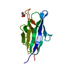
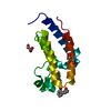
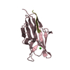
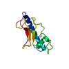
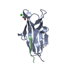
| Title | Crystal structure of the ligand-binding domain of murine SHPS-1/SIRP alpha | ||||||
 Components Components | Tyrosine-protein phosphatase non-receptor type substrate 1 | ||||||
 Keywords Keywords |  IMMUNE SYSTEM / BETA-SANDWICH / Structural Genomics / NPPSFA / National Project on Protein Structural and Functional Analyses / Alternative splicing / Glycoprotein / Immunoglobulin domain / Membrane / Phosphorylation / Polymorphism / SH3-binding / Transmembrane IMMUNE SYSTEM / BETA-SANDWICH / Structural Genomics / NPPSFA / National Project on Protein Structural and Functional Analyses / Alternative splicing / Glycoprotein / Immunoglobulin domain / Membrane / Phosphorylation / Polymorphism / SH3-binding / Transmembrane | ||||||
| Function / homology |  Function and homology information Function and homology informationSignal regulatory protein family interactions / negative regulation of I-kappaB phosphorylation / cellular response to interleukin-12 / monocyte extravasation / negative regulation of macrophage inflammatory protein 1 alpha production / granulocyte migration / negative regulation of chemokine (C-C motif) ligand 5 production / protein binding involved in heterotypic cell-cell adhesion / protein phosphorylated amino acid binding / regulation of interleukin-1 beta production ...Signal regulatory protein family interactions / negative regulation of I-kappaB phosphorylation / cellular response to interleukin-12 / monocyte extravasation / negative regulation of macrophage inflammatory protein 1 alpha production / granulocyte migration / negative regulation of chemokine (C-C motif) ligand 5 production / protein binding involved in heterotypic cell-cell adhesion / protein phosphorylated amino acid binding / regulation of interleukin-1 beta production / regulation of type II interferon production / GTPase regulator activity / Cell surface interactions at the vascular wall / positive regulation of cell-cell adhesion / cell-cell adhesion mediator activity / protein antigen binding / negative regulation of nitric oxide biosynthetic process / phagocytosis, recognition / negative regulation of interferon-beta production / negative regulation of JNK cascade / regulation of tumor necrosis factor production / regulation of nitric oxide biosynthetic process / negative regulation of phagocytosis / DAP12 interactions / regulation of interleukin-6 production / phagocytosis, engulfment / plasma membrane => GO:0005886 / negative regulation of interleukin-6 production / negative regulation of tumor necrosis factor production / negative regulation of cytokine production involved in inflammatory response / cellular response to interleukin-1 / hematopoietic progenitor cell differentiation / positive regulation of phagocytosis / cytoskeleton organization / Neutrophil degranulation / cell-matrix adhesion / protein tyrosine kinase binding / negative regulation of protein phosphorylation / actin filament organization / negative regulation of ERK1 and ERK2 cascade / negative regulation of inflammatory response / SH3 domain binding / cellular response to type II interferon / cellular response to hydrogen peroxide / cell migration / positive regulation of T cell activation / regulation of gene expression / protein phosphatase binding / cellular response to lipopolysaccharide / cell surface / plasma membraneSimilarity search - Function | ||||||
| Biological species |  Mus musculus (house mouse) Mus musculus (house mouse) | ||||||
| Method | X-RAY DIFFRACTION / SYNCHROTRON / MOLECULAR REPLACEMENT / Resolution: 1.4 Å | ||||||
 Authors Authors | Nakaishi, A. | ||||||
 Citation Citation | Journal: J.Mol.Biol. / Year: 2008 Title: Structural insight into the specific interaction between murine SHPS-1/SIRP alpha and its ligand CD47 Authors: Nakaishi, A. / Hirose, M. / Yoshimura, M. / Oneyama, C. / Saito, K. / Kuki, N. / Matsuda, M. / Honma, N. / Ohnishi, H. / Matozaki, T. / Okada, M. / Nakagawa, A. | ||||||
| History |
|
- Structure visualization
| Structure viewer | Molecule: MolmilJmol/JSmol |
|---|
- Downloads & links
-Download
| PDBx/mmCIF format | 2yz1.cif.gz | 59.8 KB | Display | PDBx/mmCIF format |
|---|---|---|---|---|
| PDB format | pdb2yz1.ent.gz | 47.6 KB | Display | PDB format |
| PDBx/mmJSON format | 2yz1.json.gz | Tree view | PDBx/mmJSON format | |
| Others |  Other downloads Other downloads |
-Validation report
| Arichive directory | https://data.pdbj.org/pub/pdb/validation_reports/yz/2yz1ftp://data.pdbj.org/pub/pdb/validation_reports/yz/2yz1 | HTTPS FTP |
|---|
-Related structure data
| Similar structure data |
|---|


-Links
 PDBj
PDBj


- Assembly
| Deposited unit | 
| ||||||||
|---|---|---|---|---|---|---|---|---|---|
| 1 | 
| ||||||||
| 2 | 
| ||||||||
| 3 |
| ||||||||
| Unit cell |
|
-Components
| #1: Protein | Mass: 13013.616 Da / Num. of mol.: 2 / Fragment: ligand-binidng domain, Ig-like V-type domain Source method: isolated from a genetically manipulated source Source: (gene. exp.) Mus musculus (house mouse) / Gene: Shps1 / Plasmid: pGEX-6P-1 / Production host:  Escherichia coli (E. coli) / Strain (production host): Rosetta-gami B / References: UniProt: P97797 Escherichia coli (E. coli) / Strain (production host): Rosetta-gami B / References: UniProt: P97797#2: Water | ChemComp-HOH / | Water Mass: 18.015 Da / Num. of mol.: 303 / Source method: isolated from a natural source / Formula: H2O Mass: 18.015 Da / Num. of mol.: 303 / Source method: isolated from a natural source / Formula: H2O |
|---|
-Experimental details
-Experiment
| Experiment | Method: X-RAY DIFFRACTION / Number of used crystals: 1 |
|---|
- Sample preparation
| Crystal | Density Matthews: 1.86 Å3/Da / Density % sol: 33.9 % |
|---|---|
| Crystal grow | Temperature: 293 K / Method: vapor diffusion, hanging drop / pH: 6.5 Details: 32% PEG 4000, 0.1M PIPES, pH 6.5, VAPOR DIFFUSION, HANGING DROP, temperature 293K |
-Data collection
| Diffraction | Mean temperature: 100 K |
|---|---|
| Diffraction source | Source: SYNCHROTRON / Site: SPring-8  / Beamline: BL44XU / Wavelength: 0.9 Å / Beamline: BL44XU / Wavelength: 0.9 Å |
| Detector | Type: Bruker DIP-6040 / Detector: CCD / Date: Apr 3, 2006 / Details: vertical focusing mirror |
| Radiation | Protocol: SINGLE WAVELENGTH / Monochromatic (M) / Laue (L): M / Scattering type: x-ray |
| Radiation wavelength | Wavelength: 0.9 Å / Relative weight: 1 |
| Reflection | Resolution: 1.4→39.75 Å / Num. obs: 39022 / % possible obs: 100 % / Observed criterion σ(F): 0 / Observed criterion σ(I): 0 / Redundancy: 7.2 % / Biso Wilson estimate: 13.2 Å2 / Rmerge(I) obs: 0.055 / Net I/σ(I): 7.3 |
| Reflection shell | Resolution: 1.4→1.48 Å / Redundancy: 7.2 % / Rmerge(I) obs: 0.34 / Num. unique all: 5624 / % possible all: 100 |
- Processing
| Software |
| ||||||||||||||||||||||||||||||||||||||||||||||||||||||||||||||||||||||||||||||||||||||||||||||||||||||||||||||||||||||||||||||||||||||||||||||||||||||||||||||||||||||||||
|---|---|---|---|---|---|---|---|---|---|---|---|---|---|---|---|---|---|---|---|---|---|---|---|---|---|---|---|---|---|---|---|---|---|---|---|---|---|---|---|---|---|---|---|---|---|---|---|---|---|---|---|---|---|---|---|---|---|---|---|---|---|---|---|---|---|---|---|---|---|---|---|---|---|---|---|---|---|---|---|---|---|---|---|---|---|---|---|---|---|---|---|---|---|---|---|---|---|---|---|---|---|---|---|---|---|---|---|---|---|---|---|---|---|---|---|---|---|---|---|---|---|---|---|---|---|---|---|---|---|---|---|---|---|---|---|---|---|---|---|---|---|---|---|---|---|---|---|---|---|---|---|---|---|---|---|---|---|---|---|---|---|---|---|---|---|---|---|---|---|---|---|
| Refinement | Method to determine structure: MOLECULAR REPLACEMENT / Resolution: 1.4→39.75 Å / Cor.coef. Fo:Fc: 0.962 / Cor.coef. Fo:Fc free: 0.957 / SU B: 0.769 / SU ML: 0.032 / Cross valid method: THROUGHOUT / ESU R: 0.067 / ESU R Free: 0.068 / Stereochemistry target values: MAXIMUM LIKELIHOOD / Details: HYDROGENS HAVE BEEN ADDED IN THE RIDING POSITIONS
| ||||||||||||||||||||||||||||||||||||||||||||||||||||||||||||||||||||||||||||||||||||||||||||||||||||||||||||||||||||||||||||||||||||||||||||||||||||||||||||||||||||||||||
| Solvent computation | Ion probe radii: 0.8 Å / Shrinkage radii: 0.8 Å / VDW probe radii: 1.4 Å / Solvent model: MASK | ||||||||||||||||||||||||||||||||||||||||||||||||||||||||||||||||||||||||||||||||||||||||||||||||||||||||||||||||||||||||||||||||||||||||||||||||||||||||||||||||||||||||||
| Displacement parameters | Biso mean: 15.984 Å2
| ||||||||||||||||||||||||||||||||||||||||||||||||||||||||||||||||||||||||||||||||||||||||||||||||||||||||||||||||||||||||||||||||||||||||||||||||||||||||||||||||||||||||||
| Refinement step | Cycle: LAST / Resolution: 1.4→39.75 Å
| ||||||||||||||||||||||||||||||||||||||||||||||||||||||||||||||||||||||||||||||||||||||||||||||||||||||||||||||||||||||||||||||||||||||||||||||||||||||||||||||||||||||||||
| Refine LS restraints |
| ||||||||||||||||||||||||||||||||||||||||||||||||||||||||||||||||||||||||||||||||||||||||||||||||||||||||||||||||||||||||||||||||||||||||||||||||||||||||||||||||||||||||||
| LS refinement shell | Resolution: 1.4→1.436 Å / Total num. of bins used: 20
|
