 Movie
Movie Controller


+ Open data

- Basic information
| Entry | Database: PDB / ID: 3jym | ||||||
|---|---|---|---|---|---|---|---|
| Title | Crystal Structure of the 3 FKBP domains of wheat FKBP73 | ||||||
 Components Components | FK506-binding protein (FKBP) from wheat | ||||||
 Keywords Keywords |  ISOMERASE / KBP- binding domain five-stranded anti-parallel -sheet and an -helix crossing this sheet / Structural Genomics / Israel Structural Proteomics Center / ISPC ISOMERASE / KBP- binding domain five-stranded anti-parallel -sheet and an -helix crossing this sheet / Structural Genomics / Israel Structural Proteomics Center / ISPC | ||||||
| Function / homology |  Function and homology information Function and homology informationchaperone-mediated protein folding / peptidylprolyl isomerase / peptidyl-prolyl cis-trans isomerase activity / calmodulin binding / cytoplasmSimilarity search - Function | ||||||
| Biological species |  Triticum aestivum (bread wheat) Triticum aestivum (bread wheat) | ||||||
| Method | X-RAY DIFFRACTION / SYNCHROTRON / PHASER / Resolution: 2.28 Å | ||||||
 Authors Authors | Dym, O. / Breiman, A. / Israel Structural Proteomics Center (ISPC) | ||||||
 Citation Citation | Journal: J.STRUCT.FUNCT.GENOM. / Year: 2010 Title: Crystal structure of the three FK506 binding protein domains of wheat FKBP73: evidence for a unique wFK73_2 domain Authors: Unger, T. / Dym, O. / Albeck, S. / Jacobovitch, Y. / Bernehim, R. / Marom, D. / Pisanty, O. / Breiman, A. | ||||||
| History |
|
- Structure visualization
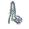
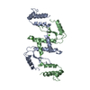
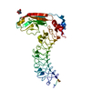
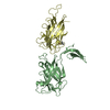
| Structure viewer | Molecule: MolmilJmol/JSmol |
|---|
- Downloads & links
-Download
| PDBx/mmCIF format | 3jym.cif.gz | 128.8 KB | Display | PDBx/mmCIF format |
|---|---|---|---|---|
| PDB format | pdb3jym.ent.gz | 100.6 KB | Display | PDB format |
| PDBx/mmJSON format | 3jym.json.gz | Tree view | PDBx/mmJSON format | |
| Others |  Other downloads Other downloads |
-Validation report
| Arichive directory | https://data.pdbj.org/pub/pdb/validation_reports/jy/3jymftp://data.pdbj.org/pub/pdb/validation_reports/jy/3jym | HTTPS FTP |
|---|
-Related structure data


-Links
 PDBj
PDBj


- Assembly
| Deposited unit | 
| ||||||||
|---|---|---|---|---|---|---|---|---|---|
| 1 | 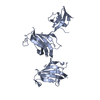
| ||||||||
| 2 | 
| ||||||||
| Unit cell |
|
-Components
| #1: Protein | Mass: 41241.625 Da / Num. of mol.: 2 Source method: isolated from a genetically manipulated source Source: (gene. exp.) Triticum aestivum (bread wheat) / Production host:  Escherichia coli (E. coli) / References: UniProt: Q43207 Escherichia coli (E. coli) / References: UniProt: Q43207 |
|---|
-Experimental details
-Experiment
| Experiment | Method: X-RAY DIFFRACTION / Number of used crystals: 1 |
|---|
- Sample preparation
| Crystal | Density Matthews: 2.42 Å3/Da / Density % sol: 49.16 % |
|---|---|
| Crystal grow | Temperature: 292 K / Method: vapor diffusion, sitting drop / pH: 6.5 Details: 0.2M NaNitrat 0.1M Bis Tris pH=6.5 20% PEG 3500, VAPOR DIFFUSION, SITTING DROP, temperature 292K |
-Data collection
| Diffraction | Mean temperature: 100 K |
|---|---|
| Diffraction source | Source: SYNCHROTRON / Site: ESRF  / Beamline: ID14-1 / Wavelength: 0.9334 Å / Beamline: ID14-1 / Wavelength: 0.9334 Å |
| Detector | Type: ADSC QUANTUM 210 / Detector: CCD / Date: Sep 5, 2007 |
| Radiation | Monochromator: Diamond / Protocol: SINGLE WAVELENGTH / Monochromatic (M) / Laue (L): M / Scattering type: x-ray |
| Radiation wavelength | Wavelength: 0.9334 Å / Relative weight: 1 |
| Reflection | Resolution: 2.28→46.63 Å / Num. obs: 36745 / % possible obs: 98.7 % / Observed criterion σ(F): 0 / Observed criterion σ(I): 0 / Redundancy: 5.2 % / Rmerge(I) obs: 0.077 / Rsym value: 0.059 / Net I/σ(I): 21 |
| Reflection shell | Resolution: 2.28→2.34 Å / Redundancy: 4.5 % / Rmerge(I) obs: 0.484 / Mean I/σ(I) obs: 3.36 / Rsym value: 0.433 / % possible all: 95.4 |
- Processing
| Software | Name: REFMAC / Version: 5.2.0019 / Classification: refinement | ||||||||||||||||||||||||||||||||||||||||||||||||||||||||||||||||||||||||||||||||||||||||||||||||||||||||||||||||||||||||||||||||||||||||||||||||||||||||||||||||||||||||||
|---|---|---|---|---|---|---|---|---|---|---|---|---|---|---|---|---|---|---|---|---|---|---|---|---|---|---|---|---|---|---|---|---|---|---|---|---|---|---|---|---|---|---|---|---|---|---|---|---|---|---|---|---|---|---|---|---|---|---|---|---|---|---|---|---|---|---|---|---|---|---|---|---|---|---|---|---|---|---|---|---|---|---|---|---|---|---|---|---|---|---|---|---|---|---|---|---|---|---|---|---|---|---|---|---|---|---|---|---|---|---|---|---|---|---|---|---|---|---|---|---|---|---|---|---|---|---|---|---|---|---|---|---|---|---|---|---|---|---|---|---|---|---|---|---|---|---|---|---|---|---|---|---|---|---|---|---|---|---|---|---|---|---|---|---|---|---|---|---|---|---|---|
| Refinement | Method to determine structure: PHASER / Resolution: 2.28→46.63 Å / Cor.coef. Fo:Fc: 0.889 / Cor.coef. Fo:Fc free: 0.828 / SU B: 8.314 / SU ML: 0.213 / Cross valid method: THROUGHOUT / σ(F): 0 / ESU R: 0.342 / ESU R Free: 0.294 / Stereochemistry target values: MAXIMUM LIKELIHOOD / Details: HYDROGENS HAVE BEEN ADDED IN THE RIDING POSITIONS
| ||||||||||||||||||||||||||||||||||||||||||||||||||||||||||||||||||||||||||||||||||||||||||||||||||||||||||||||||||||||||||||||||||||||||||||||||||||||||||||||||||||||||||
| Solvent computation | Ion probe radii: 0.8 Å / Shrinkage radii: 0.8 Å / VDW probe radii: 1.2 Å / Solvent model: MASK | ||||||||||||||||||||||||||||||||||||||||||||||||||||||||||||||||||||||||||||||||||||||||||||||||||||||||||||||||||||||||||||||||||||||||||||||||||||||||||||||||||||||||||
| Displacement parameters | Biso mean: 33.451 Å2
| ||||||||||||||||||||||||||||||||||||||||||||||||||||||||||||||||||||||||||||||||||||||||||||||||||||||||||||||||||||||||||||||||||||||||||||||||||||||||||||||||||||||||||
| Refinement step | Cycle: LAST / Resolution: 2.28→46.63 Å
| ||||||||||||||||||||||||||||||||||||||||||||||||||||||||||||||||||||||||||||||||||||||||||||||||||||||||||||||||||||||||||||||||||||||||||||||||||||||||||||||||||||||||||
| Refine LS restraints |
| ||||||||||||||||||||||||||||||||||||||||||||||||||||||||||||||||||||||||||||||||||||||||||||||||||||||||||||||||||||||||||||||||||||||||||||||||||||||||||||||||||||||||||
| LS refinement shell | Resolution: 2.279→2.338 Å / Total num. of bins used: 20
|
