 Movie
Movie Controller

[English] 日本語
 Yorodumi
- PDB-3jxe: Crystal structure of Pyrococcus horikoshii tryptophanyl-tRNA synt... -
+ Open data

- Basic information
| Entry | Database: PDB / ID: 3jxe | ||||||
|---|---|---|---|---|---|---|---|
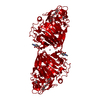
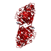
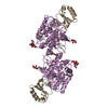
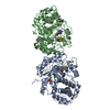
| Title | Crystal structure of Pyrococcus horikoshii tryptophanyl-tRNA synthetase in complex with TrpAMP | ||||||
 Components Components | Tryptophanyl-tRNA synthetase | ||||||
 Keywords Keywords |  LIGASE / tryptophanyl-tRNA synthetase / Adenosine Triphosphate / Rossmann fold / P. horikoshii / Aminoacyl-tRNA synthetase / ATP-binding / Cytoplasm / Nucleotide-binding / Protein biosynthesis LIGASE / tryptophanyl-tRNA synthetase / Adenosine Triphosphate / Rossmann fold / P. horikoshii / Aminoacyl-tRNA synthetase / ATP-binding / Cytoplasm / Nucleotide-binding / Protein biosynthesis | ||||||
| Function / homology |  Function and homology informationtryptophan-tRNA ligase / tryptophan-tRNA ligase activity / tryptophanyl-tRNA aminoacylation / ATP binding / cytoplasm Function and homology informationtryptophan-tRNA ligase / tryptophan-tRNA ligase activity / tryptophanyl-tRNA aminoacylation / ATP binding / cytoplasmSimilarity search - Function | ||||||
| Biological species |   Pyrococcus horikoshii (archaea) Pyrococcus horikoshii (archaea) | ||||||
| Method | X-RAY DIFFRACTION / SYNCHROTRON / MOLECULAR REPLACEMENT / Resolution: 3 Å | ||||||
 Authors Authors | Zhou, M. / Dong, X. / Zhong, C. / Shen, N. / Yang, B. / Ding, J. | ||||||
 Citation Citation | Journal: To be Published Title: Crystal structure of P. horikoshii tryptophanyl-tRNA synthetase and structure-based phylogenetic analysis suggest an archaeal origin of tryptophanyl-tRNA synthetase Authors: Zhou, M. / Dong, X. / Zhong, C. / Shen, N. / Yang, B. / Ding, J. | ||||||
| History |
|
- Structure visualization
| Structure viewer | Molecule: MolmilJmol/JSmol |
|---|
- Downloads & links
-Download
| PDBx/mmCIF format | 3jxe.cif.gz | 158.1 KB | Display | PDBx/mmCIF format |
|---|---|---|---|---|
| PDB format | pdb3jxe.ent.gz | 125.7 KB | Display | PDB format |
| PDBx/mmJSON format | 3jxe.json.gz | Tree view | PDBx/mmJSON format | |
| Others |  Other downloads Other downloads |
-Validation report
| Arichive directory | https://data.pdbj.org/pub/pdb/validation_reports/jx/3jxeftp://data.pdbj.org/pub/pdb/validation_reports/jx/3jxe | HTTPS FTP |
|---|
-Related structure data
| Similar structure data |
|---|


-Links
 PDBj
PDBj
- Assembly
| Deposited unit | 
| ||||||||
|---|---|---|---|---|---|---|---|---|---|
| 1 |
| ||||||||
| Unit cell |
|
-Components
| #1: Protein | Mass: 46202.137 Da / Num. of mol.: 2 Source method: isolated from a genetically manipulated source Source: (gene. exp.) Pyrococcus horikoshii (archaea) / Gene: PH1921, trpS / Plasmid: pET28a / Production host:  Escherichia coli (E. coli) / Strain (production host): BL21(DE3) / References: UniProt: O59584, tryptophan-tRNA ligase Escherichia coli (E. coli) / Strain (production host): BL21(DE3) / References: UniProt: O59584, tryptophan-tRNA ligase#2: Chemical |   Mass: 533.431 Da / Num. of mol.: 2 / Source method: obtained synthetically / Formula: C21H24N7O8P Mass: 533.431 Da / Num. of mol.: 2 / Source method: obtained synthetically / Formula: C21H24N7O8P#3: Chemical | Sulfate  Mass: 96.063 Da / Num. of mol.: 2 / Source method: obtained synthetically / Formula: SO4 Mass: 96.063 Da / Num. of mol.: 2 / Source method: obtained synthetically / Formula: SO4#4: Water | ChemComp-HOH / | Water Mass: 18.015 Da / Num. of mol.: 12 / Source method: isolated from a natural source / Formula: H2O Mass: 18.015 Da / Num. of mol.: 12 / Source method: isolated from a natural source / Formula: H2O |
|---|
-Experimental details
-Experiment
| Experiment | Method: X-RAY DIFFRACTION / Number of used crystals: 1 |
|---|
- Sample preparation
| Crystal | Density Matthews: 4.45 Å3/Da / Density % sol: 72.34 % |
|---|---|
| Crystal grow | Temperature: 293 K / Method: vapor diffusion, hanging drop / pH: 5.2 Details: 0.1M sodium citrate, 1.6M (NH4)2SO4, 10mM MnCl2, pH 5.2, VAPOR DIFFUSION, HANGING DROP, temperature 293K |
-Data collection
| Diffraction | Mean temperature: 100 K |
|---|---|
| Diffraction source | Source: SYNCHROTRON / Site: Photon Factory  / Beamline: BL-17A / Wavelength: 1 Å / Beamline: BL-17A / Wavelength: 1 Å |
| Detector | Type: ADSC QUANTUM 270 / Detector: CCD / Date: May 24, 2008 |
| Radiation | Protocol: SINGLE WAVELENGTH / Monochromatic (M) / Laue (L): M / Scattering type: x-ray |
| Radiation wavelength | Wavelength: 1 Å / Relative weight: 1 |
| Reflection | Resolution: 3→50 Å / Num. obs: 33131 / % possible obs: 100 % / Redundancy: 15.5 % / Biso Wilson estimate: 99.3 Å2 / Rmerge(I) obs: 0.088 / Net I/σ(I): 33.1 |
| Reflection shell | Resolution: 3→3.11 Å / Redundancy: 10.3 % / Rmerge(I) obs: 0.788 / Mean I/σ(I) obs: 2.6 / Num. unique all: 3255 / % possible all: 99.5 |
- Processing
| Software |
| ||||||||||||||||||||||||||||||||||||||||||||||||||||||||||||||||||||||||||||||||||||||||||
|---|---|---|---|---|---|---|---|---|---|---|---|---|---|---|---|---|---|---|---|---|---|---|---|---|---|---|---|---|---|---|---|---|---|---|---|---|---|---|---|---|---|---|---|---|---|---|---|---|---|---|---|---|---|---|---|---|---|---|---|---|---|---|---|---|---|---|---|---|---|---|---|---|---|---|---|---|---|---|---|---|---|---|---|---|---|---|---|---|---|---|---|
| Refinement | Method to determine structure: MOLECULAR REPLACEMENT / Resolution: 3→50 Å / Cor.coef. Fo:Fc: 0.927 / Cor.coef. Fo:Fc free: 0.905 / Occupancy max: 1 / Occupancy min: 0.7 / SU B: 15.764 / SU ML: 0.276 / Cross valid method: THROUGHOUT / σ(F): 0 / ESU R: 0.684 / ESU R Free: 0.347 / Stereochemistry target values: MAXIMUM LIKELIHOOD
| ||||||||||||||||||||||||||||||||||||||||||||||||||||||||||||||||||||||||||||||||||||||||||
| Solvent computation | Ion probe radii: 0.8 Å / Shrinkage radii: 0.8 Å / VDW probe radii: 1.2 Å / Solvent model: MASK | ||||||||||||||||||||||||||||||||||||||||||||||||||||||||||||||||||||||||||||||||||||||||||
| Displacement parameters | Biso max: 123.59 Å2 / Biso mean: 79.725 Å2 / Biso min: 41.91 Å2 | ||||||||||||||||||||||||||||||||||||||||||||||||||||||||||||||||||||||||||||||||||||||||||
| Refinement step | Cycle: LAST / Resolution: 3→50 Å
| ||||||||||||||||||||||||||||||||||||||||||||||||||||||||||||||||||||||||||||||||||||||||||
| Refine LS restraints |
| ||||||||||||||||||||||||||||||||||||||||||||||||||||||||||||||||||||||||||||||||||||||||||
| LS refinement shell | Resolution: 3→3.078 Å / Total num. of bins used: 20
|
