 Movie
Movie Controller

[English] 日本語
 Yorodumi
- PDB-3jtq: Mutations in Cephalosporin Acylase Affecting Stability and Autopr... -
+ Open data

- Basic information
| Entry | Database: PDB / ID: 3jtq | ||||||
|---|---|---|---|---|---|---|---|
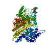
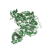
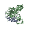
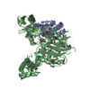
| Title | Mutations in Cephalosporin Acylase Affecting Stability and Autoproteolysis | ||||||
 Components Components | (Glutaryl 7-aminocephalosporanic acid acylase) x 2 | ||||||
 Keywords Keywords |  HYDROLASE / cephalosporin acylase / Autoproteolysis HYDROLASE / cephalosporin acylase / Autoproteolysis | ||||||
| Function / homology |  Function and homology information Function and homology informationhydrolase activity, acting on carbon-nitrogen (but not peptide) bonds, in linear amides / antibiotic biosynthetic process / periplasmic spaceSimilarity search - Function | ||||||
| Biological species |  Pseudomonas sp. (bacteria) Pseudomonas sp. (bacteria) | ||||||
| Method | X-RAY DIFFRACTION / SYNCHROTRON / MOLECULAR REPLACEMENT / Resolution: 2.2 Å | ||||||
 Authors Authors | Cho, K.J. / Kim, J.K. / Lee, J.H. / Shin, H.J. / Park, S.S. / Kim, K.H. | ||||||
 Citation Citation | Journal: Biochem.Biophys.Res.Commun. / Year: 2009 Title: Structural features of cephalosporin acylase reveal the basis of autocatalytic activation. Authors: Cho, K.J. / Kim, J.K. / Lee, J.H. / Shin, H.J. / Park, S.S. / Kim, K.H. | ||||||
| History |
|
- Structure visualization
| Structure viewer | Molecule: MolmilJmol/JSmol |
|---|
- Downloads & links
-Download
| PDBx/mmCIF format | 3jtq.cif.gz | 279.4 KB | Display | PDBx/mmCIF format |
|---|---|---|---|---|
| PDB format | pdb3jtq.ent.gz | 225.2 KB | Display | PDB format |
| PDBx/mmJSON format | 3jtq.json.gz | Tree view | PDBx/mmJSON format | |
| Others |  Other downloads Other downloads |
-Validation report
| Arichive directory | https://data.pdbj.org/pub/pdb/validation_reports/jt/3jtqftp://data.pdbj.org/pub/pdb/validation_reports/jt/3jtq | HTTPS FTP |
|---|
-Related structure data
| Related structure data |  3jtrC  2advS S: Starting model for refinement C: citing same article ( |
|---|---|
| Similar structure data |


-Links
 PDBj
PDBj


- Assembly
| Deposited unit | 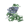
| ||||||||
|---|---|---|---|---|---|---|---|---|---|
| 1 |
| ||||||||
| Unit cell |
|
-Components
| #1: Protein | Mass: 18506.191 Da / Num. of mol.: 1 / Fragment: UNP residues 30-198 Source method: isolated from a genetically manipulated source Source: (gene. exp.) Pseudomonas sp. (bacteria) / Strain: GK16 / Plasmid: pET23d / Production host: Escherichia coli (E. coli) / Strain (production host): BL21(DE3)References: UniProt: A4ZVL3, glutaryl-7-aminocephalosporanic-acid acylase | ||||
|---|---|---|---|---|---|
| #2: Protein | Mass: 59169.777 Da / Num. of mol.: 1 / Fragment: UNP residues 199-720 / Mutation: L210N Source method: isolated from a genetically manipulated source Source: (gene. exp.) Pseudomonas sp. (bacteria) / Strain: GK16 / Plasmid: pET23d / Production host: Escherichia coli (E. coli) / Strain (production host): BL21(DE3)References: UniProt: A4ZVL3, glutaryl-7-aminocephalosporanic-acid acylase | ||||
| #3: Chemical | Glycerol  Mass: 92.094 Da / Num. of mol.: 2 / Source method: obtained synthetically / Formula: C3H8O3 Mass: 92.094 Da / Num. of mol.: 2 / Source method: obtained synthetically / Formula: C3H8O3#4: Water | ChemComp-HOH / | Water Mass: 18.015 Da / Num. of mol.: 350 / Source method: isolated from a natural source / Formula: H2O Mass: 18.015 Da / Num. of mol.: 350 / Source method: isolated from a natural source / Formula: H2OSequence details | THE DEPOSITORS | |
-Experimental details
-Experiment
| Experiment | Method: X-RAY DIFFRACTION / Number of used crystals: 1 |
|---|
- Sample preparation
| Crystal | Density Matthews: 3.37 Å3/Da / Density % sol: 63.51 % |
|---|---|
| Crystal grow | Temperature: 295 K / Method: vapor diffusion, hanging drop / pH: 6.5 Details: 0.1M Na-cacodylate (pH 6.5), 0.2M magnesium chloride, PEG 3000, VAPOR DIFFUSION, HANGING DROP, temperature 295K |
-Data collection
| Diffraction | Mean temperature: 100 K |
|---|---|
| Diffraction source | Source: SYNCHROTRON / Site: PAL/PLS  / Beamline: 4A / Wavelength: 1 Å / Beamline: 4A / Wavelength: 1 Å |
| Detector | Type: ADSC QUANTUM 210 / Detector: CCD / Date: Oct 12, 2005 |
| Radiation | Protocol: SINGLE WAVELENGTH / Monochromatic (M) / Laue (L): M / Scattering type: x-ray |
| Radiation wavelength | Wavelength: 1 Å / Relative weight: 1 |
| Reflection | Resolution: 2.2→50 Å / Num. all: 55819 / Num. obs: 53754 / % possible obs: 96.3 % / Rmerge(I) obs: 0.07 / Net I/σ(I): 32.9 |
| Reflection shell | Resolution: 2.2→2.28 Å / Rmerge(I) obs: 0.414 / Mean I/σ(I) obs: 5 / % possible all: 90.4 |
- Processing
| Software |
| |||||||||||||||||||||||||||||||||||||||||||||||||||||||||||||||||||||||||||||||||||||||||||||||||||||||||
|---|---|---|---|---|---|---|---|---|---|---|---|---|---|---|---|---|---|---|---|---|---|---|---|---|---|---|---|---|---|---|---|---|---|---|---|---|---|---|---|---|---|---|---|---|---|---|---|---|---|---|---|---|---|---|---|---|---|---|---|---|---|---|---|---|---|---|---|---|---|---|---|---|---|---|---|---|---|---|---|---|---|---|---|---|---|---|---|---|---|---|---|---|---|---|---|---|---|---|---|---|---|---|---|---|---|---|
| Refinement | Method to determine structure: MOLECULAR REPLACEMENT Starting model: PDB ENTRY 2ADV Resolution: 2.2→48.39 Å / Cor.coef. Fo:Fc: 0.939 / Cor.coef. Fo:Fc free: 0.918 / Occupancy max: 1 / Occupancy min: 1 / SU B: 12.959 / SU ML: 0.145 / Cross valid method: THROUGHOUT / σ(F): 0 / ESU R Free: 0.19 / Stereochemistry target values: MAXIMUM LIKELIHOOD / Details: HYDROGENS HAVE BEEN ADDED IN THE RIDING POSITIONS
| |||||||||||||||||||||||||||||||||||||||||||||||||||||||||||||||||||||||||||||||||||||||||||||||||||||||||
| Solvent computation | Ion probe radii: 0.8 Å / Shrinkage radii: 0.8 Å / VDW probe radii: 1.2 Å / Solvent model: MASK | |||||||||||||||||||||||||||||||||||||||||||||||||||||||||||||||||||||||||||||||||||||||||||||||||||||||||
| Displacement parameters | Biso max: 68.19 Å2 / Biso mean: 34.323 Å2 / Biso min: 15.81 Å2
| |||||||||||||||||||||||||||||||||||||||||||||||||||||||||||||||||||||||||||||||||||||||||||||||||||||||||
| Refinement step | Cycle: LAST / Resolution: 2.2→48.39 Å
| |||||||||||||||||||||||||||||||||||||||||||||||||||||||||||||||||||||||||||||||||||||||||||||||||||||||||
| Refine LS restraints |
| |||||||||||||||||||||||||||||||||||||||||||||||||||||||||||||||||||||||||||||||||||||||||||||||||||||||||
| LS refinement shell | Resolution: 2.198→2.255 Å / Total num. of bins used: 20
|
