 Movie
Movie Controller

[English] 日本語
 Yorodumi
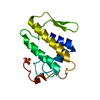
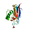
- PDB-3jql: Crystal Structure of the Complex Formed Between Phospholipase A2 ... -
+ Open data

- Basic information
| Entry | Database: PDB / ID: 3jql | ||||||
|---|---|---|---|---|---|---|---|
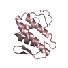
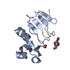
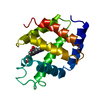
| Title | Crystal Structure of the Complex Formed Between Phospholipase A2 and a Hexapeptide Fragment of Amyloid Beta Peptide, Lys-Leu-Val-Phe-Phe-Ala at 1.2 A Resolution | ||||||
 Components Components |
| ||||||
 Keywords Keywords |  HYDROLASE / PHOSPHOLIPASE A2 / HEXAPEPTIDE / AMYLOID BETA / Disulfide bond / Lipid degradation / Metal-binding / Secreted HYDROLASE / PHOSPHOLIPASE A2 / HEXAPEPTIDE / AMYLOID BETA / Disulfide bond / Lipid degradation / Metal-binding / Secreted | ||||||
| Function / homology |  Function and homology informationphospholipase A2 / phospholipase A2 activity / arachidonic acid secretion / phospholipid metabolic process / lipid catabolic process / calcium ion binding / extracellular region Function and homology informationphospholipase A2 / phospholipase A2 activity / arachidonic acid secretion / phospholipid metabolic process / lipid catabolic process / calcium ion binding / extracellular regionSimilarity search - Function | ||||||
| Biological species |  Naja sagittifera (cobra) Naja sagittifera (cobra) | ||||||
| Method | X-RAY DIFFRACTION / MOLECULAR REPLACEMENT / Resolution: 1.2 Å | ||||||
 Authors Authors | Mirza, Z. / Vikram, G. / Singh, N. / Sinha, M. / Sharma, S. / Srinivasan, A. / Kaur, P. / Singh, T.P. | ||||||
 Citation Citation | Journal: To be Published Title: Crystal Structure of the Complex Formed Between Phospholipase A2 and a Hexapeptide Fragment of Amyloid Beta Peptide, Lys-Leu-Val-Phe-Phe-Ala at 1.2 A Resolution Authors: Mirza, Z. / Vikram, G. / Singh, N. / Sinha, M. / Sharma, S. / Srinivasan, A. / Kaur, P. / Singh, T.P. | ||||||
| History |
|
- Structure visualization
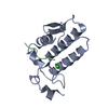
| Structure viewer | Molecule: MolmilJmol/JSmol |
|---|
- Downloads & links
-Download
| PDBx/mmCIF format | 3jql.cif.gz | 66.3 KB | Display | PDBx/mmCIF format |
|---|---|---|---|---|
| PDB format | pdb3jql.ent.gz | 48 KB | Display | PDB format |
| PDBx/mmJSON format | 3jql.json.gz | Tree view | PDBx/mmJSON format | |
| Others |  Other downloads Other downloads |
-Validation report
| Arichive directory | https://data.pdbj.org/pub/pdb/validation_reports/jq/3jqlftp://data.pdbj.org/pub/pdb/validation_reports/jq/3jql | HTTPS FTP |
|---|
-Related structure data
| Related structure data |  1mf4S S: Starting model for refinement |
|---|---|
| Similar structure data |


-Links
 PDBj
PDBj

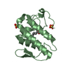
- Assembly
| Deposited unit | 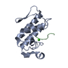
| ||||||||
|---|---|---|---|---|---|---|---|---|---|
| 1 |
| ||||||||
| Unit cell |
|
-Components
| #1: Protein | Mass: 13128.515 Da / Num. of mol.: 1 / Source method: isolated from a natural source / Source: (natural) Naja sagittifera (cobra) / References: UniProt: P60045 |
|---|---|
| #2: Protein/peptide | Amyloid beta Mass: 724.909 Da / Num. of mol.: 1 / Source method: obtained synthetically / Details: Peptide synthesis |
| #3: Chemical | ChemComp-CA /   Mass: 40.078 Da / Num. of mol.: 1 / Source method: obtained synthetically / Formula: Ca Mass: 40.078 Da / Num. of mol.: 1 / Source method: obtained synthetically / Formula: Ca |
| #4: Water | ChemComp-HOH / Water Mass: 18.015 Da / Num. of mol.: 149 / Source method: isolated from a natural source / Formula: H2O Mass: 18.015 Da / Num. of mol.: 149 / Source method: isolated from a natural source / Formula: H2O |
| Sequence details | IN CHAIN A, RESIDUE NUMBER 16 IS SIMPLY SKIPPED. THE AUTHORS BELIEVE THAT THE SEQRES IS CORRECT AND ...IN CHAIN A, RESIDUE NUMBER 16 IS SIMPLY SKIPPED. THE AUTHORS BELIEVE THAT THE SEQRES IS CORRECT AND THESE MUTATIONS ARE PRESENT IN THE DEPOSITED SEQUENCE. |
-Experimental details
-Experiment
| Experiment | Method: X-RAY DIFFRACTION / Number of used crystals: 1 |
|---|
- Sample preparation
| Crystal | Density Matthews: 2.04 Å3/Da / Density % sol: 39.83 % |
|---|---|
| Crystal grow | Temperature: 290 K / Method: vapor diffusion, hanging drop / pH: 6 Details: calcium chloride, sodium phosphate, PH6.0, VAPOR DIFFUSION, HANGING DROP, temperature 290K |
-Data collection
| Diffraction | Mean temperature: 300 K |
|---|---|
| Diffraction source | Source: ROTATING ANODE / Type: RIGAKU RU300 / Wavelength: 1.5418 Å |
| Detector | Type: MARRESEARCH / Detector: IMAGE PLATE / Date: Mar 30, 2009 / Details: MIRROR |
| Radiation | Monochromator: GRAPHITE / Protocol: SINGLE WAVELENGTH / Monochromatic (M) / Laue (L): M / Scattering type: x-ray |
| Radiation wavelength | Wavelength: 1.5418 Å / Relative weight: 1 |
| Reflection | Resolution: 1.2→42 Å / Num. all: 34924 / Num. obs: 34924 / % possible obs: 95.9 % / Observed criterion σ(F): 0 / Observed criterion σ(I): 0 / Rsym value: 0.036 / Net I/σ(I): 7.9 |
| Reflection shell | Resolution: 1.2→1.22 Å / Mean I/σ(I) obs: 2.5 / Rsym value: 0.122 / % possible all: 94.6 |
- Processing
| Software |
| ||||||||||||||||||||||||||||||||||||||||||||||||||||||||||||||||||||||||||||||||||||||||||||||||||||||||||||||
|---|---|---|---|---|---|---|---|---|---|---|---|---|---|---|---|---|---|---|---|---|---|---|---|---|---|---|---|---|---|---|---|---|---|---|---|---|---|---|---|---|---|---|---|---|---|---|---|---|---|---|---|---|---|---|---|---|---|---|---|---|---|---|---|---|---|---|---|---|---|---|---|---|---|---|---|---|---|---|---|---|---|---|---|---|---|---|---|---|---|---|---|---|---|---|---|---|---|---|---|---|---|---|---|---|---|---|---|---|---|---|---|
| Refinement | Method to determine structure: MOLECULAR REPLACEMENT Starting model: PDB ENTRY 1MF4 Resolution: 1.2→42 Å / Cor.coef. Fo:Fc: 0.962 / Cor.coef. Fo:Fc free: 0.961 / SU B: 1.294 / SU ML: 0.027 / Cross valid method: THROUGHOUT / σ(F): 0 / σ(I): 0 / ESU R: 0.052 / ESU R Free: 0.047 / Stereochemistry target values: MAXIMUM LIKELIHOOD / Details: HYDROGENS HAVE BEEN ADDED IN THE RIDING POSITIONS
| ||||||||||||||||||||||||||||||||||||||||||||||||||||||||||||||||||||||||||||||||||||||||||||||||||||||||||||||
| Solvent computation | Ion probe radii: 0.8 Å / Shrinkage radii: 0.8 Å / VDW probe radii: 1.2 Å / Solvent model: MASK | ||||||||||||||||||||||||||||||||||||||||||||||||||||||||||||||||||||||||||||||||||||||||||||||||||||||||||||||
| Displacement parameters | Biso mean: 23.326 Å2
| ||||||||||||||||||||||||||||||||||||||||||||||||||||||||||||||||||||||||||||||||||||||||||||||||||||||||||||||
| Refinement step | Cycle: LAST / Resolution: 1.2→42 Å
| ||||||||||||||||||||||||||||||||||||||||||||||||||||||||||||||||||||||||||||||||||||||||||||||||||||||||||||||
| Refine LS restraints |
| ||||||||||||||||||||||||||||||||||||||||||||||||||||||||||||||||||||||||||||||||||||||||||||||||||||||||||||||
| LS refinement shell | Resolution: 1.199→1.23 Å / Total num. of bins used: 20
|
