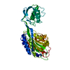
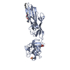
 Movie
Movie Controller


+ Open data

- Basic information
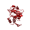
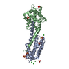
| Entry | Database: PDB / ID: 3icu | ||||||
|---|---|---|---|---|---|---|---|
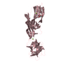
| Title | Protease-associated domain of the E3 ligase grail | ||||||
 Components Components | E3 ubiquitin-protein ligase RNF128 | ||||||
 Keywords Keywords |  LIGASE / E3 LIGASE / ENERGY / PA DOMAIN / TRANSMEMBRANE / PROTEIN TURNOVER / UBL CONJUGATION PATHWAY / GLYCOPROTEIN / STRUCTURAL GENOMICS CONSORTIUM / SGC / Membrane / Metal-binding / Zinc-finger LIGASE / E3 LIGASE / ENERGY / PA DOMAIN / TRANSMEMBRANE / PROTEIN TURNOVER / UBL CONJUGATION PATHWAY / GLYCOPROTEIN / STRUCTURAL GENOMICS CONSORTIUM / SGC / Membrane / Metal-binding / Zinc-finger | ||||||
| Function / homology |  Function and homology information Function and homology informationnegative regulation of cytokine production => GO:0001818 / positive regulation of protein catabolic process in the vacuole / protein localization to lysosome / protein deubiquitination / RING-type E3 ubiquitin transferase / regulation of protein stability / ubiquitin protein ligase activity / Ovarian tumor domain proteases / late endosome / ubiquitin-dependent protein catabolic process ...negative regulation of cytokine production => GO:0001818 / positive regulation of protein catabolic process in the vacuole / protein localization to lysosome / protein deubiquitination / RING-type E3 ubiquitin transferase / regulation of protein stability / ubiquitin protein ligase activity / Ovarian tumor domain proteases / late endosome / ubiquitin-dependent protein catabolic process / membrane => GO:0016020 / cytoskeleton / Ub-specific processing proteases / perinuclear region of cytoplasm / Golgi apparatus / endoplasmic reticulum / metal ion binding / cytosol / cytoplasmSimilarity search - Function | ||||||
| Biological species |  Homo sapiens (human) Homo sapiens (human) | ||||||
| Method | X-RAY DIFFRACTION / MOLECULAR REPLACEMENT / Resolution: 2.1 Å | ||||||
 Authors Authors | Walker, J.R. / Yermekbayeva, L. / Seitova, A. / Weigelt, J. / Bountra, C. / Arrowsmith, C.H. / Edwards, A.M. / Bochkarev, A. / Dhe-Paganon, S. / Structural Genomics Consortium (SGC) | ||||||
 Citation Citation | Journal: To be Published Title: PA Domain of the E3 Ligase Grail Authors: Walker, J.R. / Yermekbayeva, L. / Seitova, A. / Weigelt, J. / Bountra, C. / Arrowsmith, C.H. / Edwards, A.M. / Bochkarev, A. / Dhe-Paganon, S. | ||||||
| History |
|
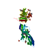
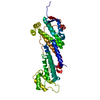
- Structure visualization
| Structure viewer | Molecule: MolmilJmol/JSmol |
|---|
- Downloads & links
-Download
| PDBx/mmCIF format | 3icu.cif.gz | 87.9 KB | Display | PDBx/mmCIF format |
|---|---|---|---|---|
| PDB format | pdb3icu.ent.gz | 65.8 KB | Display | PDB format |
| PDBx/mmJSON format | 3icu.json.gz | Tree view | PDBx/mmJSON format | |
| Others |  Other downloads Other downloads |
-Validation report
| Arichive directory | https://data.pdbj.org/pub/pdb/validation_reports/ic/3icuftp://data.pdbj.org/pub/pdb/validation_reports/ic/3icu | HTTPS FTP |
|---|
-Related structure data


-Links
 PDBj
PDBj


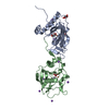
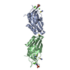
- Assembly
| Deposited unit | 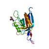
| ||||||||
|---|---|---|---|---|---|---|---|---|---|
| 1 | 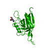
| ||||||||
| Unit cell |
| ||||||||
| Components on special symmetry positions |
|
-Components
| #1: Protein | Mass: 21051.746 Da / Num. of mol.: 1 / Fragment: UNP residues 38-204 Source method: isolated from a genetically manipulated source Source: (gene. exp.) Homo sapiens (human) / Gene: GRAIL, RNF128 / Plasmid: PFHMSP-LIC-N / Production host:   Spodoptera frugiperda (fall armyworm) / Strain (production host): SF9 Spodoptera frugiperda (fall armyworm) / Strain (production host): SF9References: UniProt: Q8TEB7, Ligases; Forming carbon-nitrogen bonds; Acid-amino-acid ligases (peptide synthases) |
|---|---|
| #2: Sugar | ChemComp-NAG / N-Acetylglucosamine  Type: D-saccharide, beta linking / Mass: 221.208 Da / Num. of mol.: 1 Type: D-saccharide, beta linking / Mass: 221.208 Da / Num. of mol.: 1Source method: isolated from a genetically manipulated source Formula: C8H15NO6 |
| #3: Water | ChemComp-HOH / Water Mass: 18.015 Da / Num. of mol.: 142 / Source method: isolated from a natural source / Formula: H2O Mass: 18.015 Da / Num. of mol.: 142 / Source method: isolated from a natural source / Formula: H2O |
-Experimental details
-Experiment
| Experiment | Method: X-RAY DIFFRACTION / Number of used crystals: 1 |
|---|
- Sample preparation
| Crystal | Density Matthews: 3.58 Å3/Da / Density % sol: 65.35 % |
|---|---|
| Crystal grow | Temperature: 290.9 K / Method: vapor diffusion, sitting drop / pH: 8 Details: 20% PEG 3350, 0.2 M NaH2PO4. PH 8.0, VAPOR DIFFUSION, SITTING DROP, TEMPERATURE 290.9K |
-Data collection
| Diffraction | Mean temperature: 100 K |
|---|---|
| Diffraction source | Source: ROTATING ANODE / Type: RIGAKU FR-E SUPERBRIGHT / Wavelength: 1.54178 / Wavelength: 1.54178 Å |
| Detector | Type: RIGAKU RAXIS IV / Detector: IMAGE PLATE / Date: Jun 25, 2009 |
| Radiation | Monochromator: GRAPHITE / Protocol: SINGLE WAVELENGTH / Monochromatic (M) / Laue (L): M / Scattering type: x-ray |
| Radiation wavelength | Wavelength: 1.54178 Å / Relative weight: 1 |
| Reflection | Resolution: 2.1→25 Å / Num. all: 18428 / Num. obs: 18428 / % possible obs: 100 % / Observed criterion σ(F): 0 / Observed criterion σ(I): -3 / Redundancy: 11.6 % / Rsym value: 0.101 / Net I/σ(I): 30.7647 |
| Reflection shell | Resolution: 2.1→2.18 Å / Redundancy: 10.2 % / Mean I/σ(I) obs: 3.633 / Num. unique all: 1804 / Rsym value: 0.748 / % possible all: 99.9 |
- Processing
| Software |
| |||||||||||||||||||||||||||||||||||||||||||||||||||||||||||||||||
|---|---|---|---|---|---|---|---|---|---|---|---|---|---|---|---|---|---|---|---|---|---|---|---|---|---|---|---|---|---|---|---|---|---|---|---|---|---|---|---|---|---|---|---|---|---|---|---|---|---|---|---|---|---|---|---|---|---|---|---|---|---|---|---|---|---|---|
| Refinement | Method to determine structure: MOLECULAR REPLACEMENT Starting model: PDB ENTRY 2EK8, 1XF1 Resolution: 2.1→25 Å / Cor.coef. Fo:Fc: 0.948 / Cor.coef. Fo:Fc free: 0.935 / SU B: 7.377 / SU ML: 0.089 / Cross valid method: THROUGHOUT / ESU R: 0.15 / ESU R Free: 0.136 / Stereochemistry target values: MAXIMUM LIKELIHOOD Details: HYDROGENS HAVE BEEN ADDED IN THE RIDING POSITIONS U VALUES: WITH TLS ADDED
| |||||||||||||||||||||||||||||||||||||||||||||||||||||||||||||||||
| Solvent computation | Ion probe radii: 0.8 Å / Shrinkage radii: 0.8 Å / VDW probe radii: 1.4 Å / Solvent model: MASK | |||||||||||||||||||||||||||||||||||||||||||||||||||||||||||||||||
| Displacement parameters | Biso mean: 32.997 Å2
| |||||||||||||||||||||||||||||||||||||||||||||||||||||||||||||||||
| Refinement step | Cycle: LAST / Resolution: 2.1→25 Å
| |||||||||||||||||||||||||||||||||||||||||||||||||||||||||||||||||
| Refine LS restraints |
| |||||||||||||||||||||||||||||||||||||||||||||||||||||||||||||||||
| LS refinement shell | Resolution: 2.1→2.154 Å / Total num. of bins used: 20
| |||||||||||||||||||||||||||||||||||||||||||||||||||||||||||||||||
| Refinement TLS params. | Method: refined / Origin x: 32.9688 Å / Origin y: 14.9519 Å / Origin z: 16.7889 Å
|
