 Movie
Movie Controller

[English] 日本語
 Yorodumi
- PDB-3ia8: The structure of the C-terminal heme nitrobindin domain of THAP d... -
+ Open data

- Basic information
| Entry | Database: PDB / ID: 3ia8 | ||||||
|---|---|---|---|---|---|---|---|
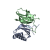
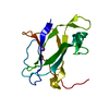
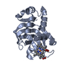
| Title | The structure of the C-terminal heme nitrobindin domain of THAP domain-containing protein 4 from Homo sapiens | ||||||
 Components Components | THAP domain-containing protein 4 | ||||||
 Keywords Keywords | METAL BINDING PROTEIN /  beta barrel / STRUCTURAL GENOMICS / PROTEIN STRUCTURE INITIATIVE / PSI / CENTER FOR EUKARYOTIC STRUCTURAL GENOMICS / CESG / Heme / THAP domain / THAP4 / DNA-binding / Metal-binding / Phosphoprotein / Zinc-finger beta barrel / STRUCTURAL GENOMICS / PROTEIN STRUCTURE INITIATIVE / PSI / CENTER FOR EUKARYOTIC STRUCTURAL GENOMICS / CESG / Heme / THAP domain / THAP4 / DNA-binding / Metal-binding / Phosphoprotein / Zinc-finger | ||||||
| Function / homology |  Function and homology information Function and homology informationperoxynitrite isomerase activity / Isomerases; Other isomerases / nitrate metabolic process / tyrosine metabolic process / chromatin => GO:0000785 / nitric oxide binding / DNA-binding transcription factor activity, RNA polymerase II-specific / heme binding / DNA binding / identical protein binding / metal ion bindingSimilarity search - Function | ||||||
| Biological species |  Homo sapiens (human) Homo sapiens (human) | ||||||
| Method | X-RAY DIFFRACTION / SYNCHROTRON / SAD / Resolution: 1.792 Å | ||||||
 Authors Authors | Bianchetti, C.M. / Bingman, C.A. / Phillips Jr., G.N. / Center for Eukaryotic Structural Genomics (CESG) | ||||||
 Citation Citation | Journal: Proteins / Year: 2011 Title: Structure of the C-terminal heme-binding domain of THAP domain containing protein 4 from Homo sapiens. Authors: Bianchetti, C.M. / Bingman, C.A. / Phillips, G.N. | ||||||
| History |
|
- Structure visualization
| Structure viewer | Molecule: MolmilJmol/JSmol |
|---|
- Downloads & links
-Download
| PDBx/mmCIF format | 3ia8.cif.gz | 83.8 KB | Display | PDBx/mmCIF format |
|---|---|---|---|---|
| PDB format | pdb3ia8.ent.gz | 67 KB | Display | PDB format |
| PDBx/mmJSON format | 3ia8.json.gz | Tree view | PDBx/mmJSON format | |
| Others |  Other downloads Other downloads |
-Validation report
| Arichive directory | https://data.pdbj.org/pub/pdb/validation_reports/ia/3ia8ftp://data.pdbj.org/pub/pdb/validation_reports/ia/3ia8 | HTTPS FTP |
|---|
-Related structure data
| Similar structure data | |
|---|---|
| Other databases |


-Links
 PDBj
PDBj

- Assembly
| Deposited unit | 
| ||||||||
|---|---|---|---|---|---|---|---|---|---|
| 1 | 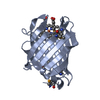
| ||||||||
| 2 | 
| ||||||||
| 3 |
| ||||||||
| Unit cell |
|
-Components
| #1: Protein | Mass: 18671.322 Da / Num. of mol.: 2 / Fragment: sequence database residues 415-577 Source method: isolated from a genetically manipulated source Source: (gene. exp.) Homo sapiens (human) / Gene: THAP4, CGI-36, PP238 / Plasmid: PVP68K / Production host:  Escherichia coli (E. coli) / Strain (production host): B834 P(RARE2) / References: UniProt: Q8WY91 Escherichia coli (E. coli) / Strain (production host): B834 P(RARE2) / References: UniProt: Q8WY91#2: Chemical | Heme B  Mass: 616.487 Da / Num. of mol.: 2 / Source method: obtained synthetically / Formula: C34H32FeN4O4 Mass: 616.487 Da / Num. of mol.: 2 / Source method: obtained synthetically / Formula: C34H32FeN4O4#3: Water | ChemComp-HOH / | Water Mass: 18.015 Da / Num. of mol.: 272 / Source method: isolated from a natural source / Formula: H2O Mass: 18.015 Da / Num. of mol.: 272 / Source method: isolated from a natural source / Formula: H2O |
|---|
-Experimental details
-Experiment
| Experiment | Method: X-RAY DIFFRACTION / Number of used crystals: 1 |
|---|
- Sample preparation
| Crystal | Density Matthews: 2.16 Å3/Da / Density % sol: 49.54 % |
|---|---|
| Crystal grow | Temperature: 277 K / Method: vapor diffusion, hanging drop / pH: 7.5 Details: Protein Solution (10 mg/ml MSE protein, 0.05 M NaCl, 0.0003 M TCEP, 0.005 M HEPES pH 5) mixed in a 1:1 ratio with the Well Solution (55% Polypropylene glycol 400, 0.1 M Triethanolamine pH 7. ...Details: Protein Solution (10 mg/ml MSE protein, 0.05 M NaCl, 0.0003 M TCEP, 0.005 M HEPES pH 5) mixed in a 1:1 ratio with the Well Solution (55% Polypropylene glycol 400, 0.1 M Triethanolamine pH 7.5). Cryoprotected with 55% Polypropylene glycol 400, 0.1 M Triethanolamine pH 7.5, vapor diffusion, hanging drop, temperature 277K |
-Data collection
| Diffraction | Mean temperature: 100 K | ||||||||||||||||||||||||||||||||||||||||||||||||||||||||||||||||||||||||||||||||||||||||||||||||||||||||||||||||
|---|---|---|---|---|---|---|---|---|---|---|---|---|---|---|---|---|---|---|---|---|---|---|---|---|---|---|---|---|---|---|---|---|---|---|---|---|---|---|---|---|---|---|---|---|---|---|---|---|---|---|---|---|---|---|---|---|---|---|---|---|---|---|---|---|---|---|---|---|---|---|---|---|---|---|---|---|---|---|---|---|---|---|---|---|---|---|---|---|---|---|---|---|---|---|---|---|---|---|---|---|---|---|---|---|---|---|---|---|---|---|---|---|---|
| Diffraction source | Source: SYNCHROTRON / Site: APS  / Beamline: 21-ID-G / Wavelength: 0.97857 Å / Beamline: 21-ID-G / Wavelength: 0.97857 Å | ||||||||||||||||||||||||||||||||||||||||||||||||||||||||||||||||||||||||||||||||||||||||||||||||||||||||||||||||
| Detector | Type: MARMOSAIC 300 mm CCD / Detector: CCD / Date: Jun 8, 2009 / Details: mirrors and beryllium lenses | ||||||||||||||||||||||||||||||||||||||||||||||||||||||||||||||||||||||||||||||||||||||||||||||||||||||||||||||||
| Radiation | Monochromator: C(111) / Protocol: SINGLE WAVELENGTH / Monochromatic (M) / Laue (L): M / Scattering type: x-ray | ||||||||||||||||||||||||||||||||||||||||||||||||||||||||||||||||||||||||||||||||||||||||||||||||||||||||||||||||
| Radiation wavelength | Wavelength: 0.97857 Å / Relative weight: 1 | ||||||||||||||||||||||||||||||||||||||||||||||||||||||||||||||||||||||||||||||||||||||||||||||||||||||||||||||||
| Reflection | Redundancy: 5.8 % / Av σ(I) over netI: 18.16 / Number: 173743 / Rmerge(I) obs: 0.093 / Χ2: 1.18 / D res high: 1.8 Å / D res low: 50 Å / Num. obs: 29918 / % possible obs: 95.9 | ||||||||||||||||||||||||||||||||||||||||||||||||||||||||||||||||||||||||||||||||||||||||||||||||||||||||||||||||
| Diffraction reflection shell |
| ||||||||||||||||||||||||||||||||||||||||||||||||||||||||||||||||||||||||||||||||||||||||||||||||||||||||||||||||
| Reflection | Resolution: 1.79→50 Å / Num. obs: 29918 / % possible obs: 95.9 % / Redundancy: 5.8 % / Biso Wilson estimate: 19.36 Å2 / Rmerge(I) obs: 0.093 / Χ2: 1.182 / Net I/σ(I): 9.5 | ||||||||||||||||||||||||||||||||||||||||||||||||||||||||||||||||||||||||||||||||||||||||||||||||||||||||||||||||
| Reflection shell |
|
-Phasing
| Phasing | Method: SAD | ||||||||||||||||||||||||||||||||||||||||||||||||||||||||||||||||||||||||||||||||||||||||||||||||||||||||||||||||||||||||||||||||||||||||||||||||||||||||||||||||||||||||||||||||||||||||||||||||||||||||||||||||||||||||||||||||||||||||||||||||||||||
|---|---|---|---|---|---|---|---|---|---|---|---|---|---|---|---|---|---|---|---|---|---|---|---|---|---|---|---|---|---|---|---|---|---|---|---|---|---|---|---|---|---|---|---|---|---|---|---|---|---|---|---|---|---|---|---|---|---|---|---|---|---|---|---|---|---|---|---|---|---|---|---|---|---|---|---|---|---|---|---|---|---|---|---|---|---|---|---|---|---|---|---|---|---|---|---|---|---|---|---|---|---|---|---|---|---|---|---|---|---|---|---|---|---|---|---|---|---|---|---|---|---|---|---|---|---|---|---|---|---|---|---|---|---|---|---|---|---|---|---|---|---|---|---|---|---|---|---|---|---|---|---|---|---|---|---|---|---|---|---|---|---|---|---|---|---|---|---|---|---|---|---|---|---|---|---|---|---|---|---|---|---|---|---|---|---|---|---|---|---|---|---|---|---|---|---|---|---|---|---|---|---|---|---|---|---|---|---|---|---|---|---|---|---|---|---|---|---|---|---|---|---|---|---|---|---|---|---|---|---|---|---|---|---|---|---|---|---|---|---|---|---|---|---|---|---|---|---|
| Phasing MAD set |
| ||||||||||||||||||||||||||||||||||||||||||||||||||||||||||||||||||||||||||||||||||||||||||||||||||||||||||||||||||||||||||||||||||||||||||||||||||||||||||||||||||||||||||||||||||||||||||||||||||||||||||||||||||||||||||||||||||||||||||||||||||||||
| Phasing MAD set shell |
| ||||||||||||||||||||||||||||||||||||||||||||||||||||||||||||||||||||||||||||||||||||||||||||||||||||||||||||||||||||||||||||||||||||||||||||||||||||||||||||||||||||||||||||||||||||||||||||||||||||||||||||||||||||||||||||||||||||||||||||||||||||||
| Phasing MAD set site |
| ||||||||||||||||||||||||||||||||||||||||||||||||||||||||||||||||||||||||||||||||||||||||||||||||||||||||||||||||||||||||||||||||||||||||||||||||||||||||||||||||||||||||||||||||||||||||||||||||||||||||||||||||||||||||||||||||||||||||||||||||||||||
| Phasing dm | Method: Solvent flattening and Histogram matching / Reflection: 29829 | ||||||||||||||||||||||||||||||||||||||||||||||||||||||||||||||||||||||||||||||||||||||||||||||||||||||||||||||||||||||||||||||||||||||||||||||||||||||||||||||||||||||||||||||||||||||||||||||||||||||||||||||||||||||||||||||||||||||||||||||||||||||
| Phasing dm shell |
|
- Processing
| Software |
| ||||||||||||||||||||||||||||||||||||||||||||||||||||||||||||||||||||||||||||||||||||
|---|---|---|---|---|---|---|---|---|---|---|---|---|---|---|---|---|---|---|---|---|---|---|---|---|---|---|---|---|---|---|---|---|---|---|---|---|---|---|---|---|---|---|---|---|---|---|---|---|---|---|---|---|---|---|---|---|---|---|---|---|---|---|---|---|---|---|---|---|---|---|---|---|---|---|---|---|---|---|---|---|---|---|---|---|---|
| Refinement | Method to determine structure: SAD / Resolution: 1.792→35.113 Å / Occupancy max: 1 / Occupancy min: 0.29 / SU ML: 0.14 / σ(F): 1.34 / Phase error: 20.05 / Stereochemistry target values: ML
| ||||||||||||||||||||||||||||||||||||||||||||||||||||||||||||||||||||||||||||||||||||
| Solvent computation | Shrinkage radii: 0.9 Å / VDW probe radii: 1.11 Å / Solvent model: FLAT BULK SOLVENT MODEL / Bsol: 49.54 Å2 / ksol: 0.349 e/Å3 | ||||||||||||||||||||||||||||||||||||||||||||||||||||||||||||||||||||||||||||||||||||
| Displacement parameters | Biso max: 73.45 Å2 / Biso mean: 24.305 Å2 / Biso min: 11.87 Å2
| ||||||||||||||||||||||||||||||||||||||||||||||||||||||||||||||||||||||||||||||||||||
| Refinement step | Cycle: LAST / Resolution: 1.792→35.113 Å
| ||||||||||||||||||||||||||||||||||||||||||||||||||||||||||||||||||||||||||||||||||||
| Refine LS restraints |
| ||||||||||||||||||||||||||||||||||||||||||||||||||||||||||||||||||||||||||||||||||||
| LS refinement shell | Refine-ID: X-RAY DIFFRACTION / Total num. of bins used: 11
|
