 Movie
Movie Controller

[English] 日本語
 Yorodumi
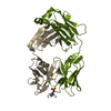
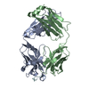
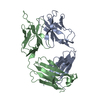
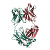
- PDB-3i9g: Crystal structure of the LT1009 (SONEPCIZUMAB) antibody Fab fragm... -
+ Open data

- Basic information
| Entry | Database: PDB / ID: 3i9g | ||||||
|---|---|---|---|---|---|---|---|
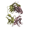
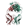
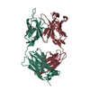
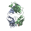
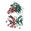
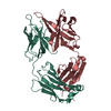
| Title | Crystal structure of the LT1009 (SONEPCIZUMAB) antibody Fab fragment in complex with sphingosine-1-phosphate | ||||||
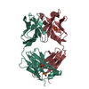
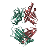
 Components Components | (Sonepcizumab antibody Fab fragment, ...) x 2 | ||||||
 Keywords Keywords |  IMMUNE SYSTEM / Antibody / Fab / Sphingosine-1-phosphate / Calcium / Immunoglobin / IgG IMMUNE SYSTEM / Antibody / Fab / Sphingosine-1-phosphate / Calcium / Immunoglobin / IgG | ||||||
| Function / homology | Immunoglobulins / Immunoglobulin-like / Sandwich / Mainly Beta / Chem-S1P Function and homology information Function and homology information | ||||||
| Biological species |  Mus musculus (house mouse) Mus musculus (house mouse) | ||||||
| Method | X-RAY DIFFRACTION / SYNCHROTRON / MOLECULAR REPLACEMENT / molecular replacement / Resolution: 1.9 Å | ||||||
 Authors Authors | Huxford, T. | ||||||
 Citation Citation | Journal: Proc.Natl.Acad.Sci.USA / Year: 2009 Title: The crystal structure of sphingosine-1-phosphate in complex with a Fab fragment reveals metal bridging of an antibody and its antigen. Authors: Wojciak, J.M. / Zhu, N. / Schuerenberg, K.T. / Moreno, K. / Shestowsky, W.S. / Hiraiwa, M. / Sabbadini, R. / Huxford, T. #1: Journal: J.Lipid Res. / Year: 2009 Title: Production and characterization of monoclonal anti-sphingosine-1-phosphate antibodies Authors: O'Brien, N. / Jones, S.T. / Williams, D.G. / Cunningham, H.B. / Moreno, K. / Visentin, B. / Gentile, A. / Vekich, J. / Shestowsky, W. / Hiraiwa, M. / Matteo, R. / Cavalli, A. / Grotjahn, D. ...Authors: O'Brien, N. / Jones, S.T. / Williams, D.G. / Cunningham, H.B. / Moreno, K. / Visentin, B. / Gentile, A. / Vekich, J. / Shestowsky, W. / Hiraiwa, M. / Matteo, R. / Cavalli, A. / Grotjahn, D. / Grant, M. / Hansen, G. / Campbell, M.A. / Sabbadini, R. | ||||||
| History |
|
- Structure visualization
| Structure viewer | Molecule: MolmilJmol/JSmol |
|---|
- Downloads & links
-Download
| PDBx/mmCIF format | 3i9g.cif.gz | 101.9 KB | Display | PDBx/mmCIF format |
|---|---|---|---|---|
| PDB format | pdb3i9g.ent.gz | 81.3 KB | Display | PDB format |
| PDBx/mmJSON format | 3i9g.json.gz | Tree view | PDBx/mmJSON format | |
| Others |  Other downloads Other downloads |
-Validation report
| Arichive directory | https://data.pdbj.org/pub/pdb/validation_reports/i9/3i9gftp://data.pdbj.org/pub/pdb/validation_reports/i9/3i9g | HTTPS FTP |
|---|
-Related structure data
| Similar structure data |
|---|


-Links
 PDBj
PDBj

- Assembly
| Deposited unit | 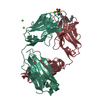
| ||||||||
|---|---|---|---|---|---|---|---|---|---|
| 1 |
| ||||||||
| Unit cell |
|
-Components
-Antibody , 2 types, 2 molecules HL
| #1: Antibody | Mass: 24027.127 Da / Num. of mol.: 1 / Fragment: Fab Fragment Source method: isolated from a genetically manipulated source Details: Humanized mouse IgG1K expressed from a stable transfected CHO cell line Source: (gene. exp.) Mus musculus (house mouse) / Gene: IgG1K / Cell line (production host): CHO cells / Production host:  Cricetulus griseus (Chinese hamster) Cricetulus griseus (Chinese hamster) |
|---|---|
| #2: Antibody | Mass: 23442.965 Da / Num. of mol.: 1 / Fragment: Fab Fragment Source method: isolated from a genetically manipulated source Details: Humanized mouse IgG1K expressed from a stable transfected CHO cell line Source: (gene. exp.) Mus musculus (house mouse) / Cell line (production host): CHO cells / Production host: Cricetulus griseus (Chinese hamster) |
-Non-polymers , 4 types, 291 molecules 

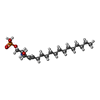

| #3: Chemical |  Mass: 40.078 Da / Num. of mol.: 2 / Source method: obtained synthetically / Formula: Ca Mass: 40.078 Da / Num. of mol.: 2 / Source method: obtained synthetically / Formula: Ca#4: Chemical | ChemComp-MG /  Mass: 24.305 Da / Num. of mol.: 5 / Source method: obtained synthetically / Formula: Mg Mass: 24.305 Da / Num. of mol.: 5 / Source method: obtained synthetically / Formula: Mg#5: Chemical | ChemComp-S1P / ( | Sphingosine-1-phosphate Mass: 379.472 Da / Num. of mol.: 1 / Source method: obtained synthetically / Formula: C18H38NO5P Mass: 379.472 Da / Num. of mol.: 1 / Source method: obtained synthetically / Formula: C18H38NO5P#6: Water | ChemComp-HOH / | WaterMass: 18.015 Da / Num. of mol.: 283 / Source method: isolated from a natural source / Formula: H2O |
|---|
-Experimental details
-Experiment
| Experiment | Method: X-RAY DIFFRACTION / Number of used crystals: 1 |
|---|
- Sample preparation
| Crystal | Density Matthews: 3.42 Å3/Da / Density % sol: 64.04 % |
|---|---|
| Crystal grow | Temperature: 291.15 K / Method: vapor diffusion, hanging drop / pH: 6 Details: 1 microliter of 12 mg/mL S1P:Fab complex was mixed with 1 microliter and sealed over 1 mL of reservoir solution containing 22% (w/v) polyethylene glycol 3350, 100 mM MgSO4, 100 mM sodium ...Details: 1 microliter of 12 mg/mL S1P:Fab complex was mixed with 1 microliter and sealed over 1 mL of reservoir solution containing 22% (w/v) polyethylene glycol 3350, 100 mM MgSO4, 100 mM sodium citrate (pH 6.0) and 10% (v/v) ethylene glycol., VAPOR DIFFUSION, HANGING DROP, temperature 291.15K |
-Data collection
| Diffraction | Mean temperature: 100 K |
|---|---|
| Diffraction source | Source: SYNCHROTRON / Site: ALS  / Beamline: 5.0.1 / Wavelength: 0.97741 Å / Beamline: 5.0.1 / Wavelength: 0.97741 Å |
| Detector | Type: ADSC QUANTUM 210 / Detector: CCD / Date: Aug 21, 2008 |
| Radiation | Monochromator: Single crystal, cylindrically bent, Si(220) / Protocol: SINGLE WAVELENGTH / Monochromatic (M) / Laue (L): M / Scattering type: x-ray |
| Radiation wavelength | Wavelength: 0.97741 Å / Relative weight: 1 |
| Reflection | Resolution: 1.9→50 Å / Num. all: 52056 / Num. obs: 50494 / % possible obs: 97 % / Observed criterion σ(I): 3.8 / Redundancy: 5.1 % / Rsym value: 0.055 / Net I/σ(I): 26.4 |
| Reflection shell | Resolution: 1.9→1.97 Å / Redundancy: 3.3 % / Mean I/σ(I) obs: 2.18 / Num. unique all: 3940 / Rsym value: 0.409 / % possible all: 76.8 |
-Phasing
| Phasing | Method: molecular replacement |
|---|
- Processing
| Software |
| |||||||||||||||||||||||||||||||||||||||||||||||||||||||||||||||||||||||||||||||||||||||||||||||
|---|---|---|---|---|---|---|---|---|---|---|---|---|---|---|---|---|---|---|---|---|---|---|---|---|---|---|---|---|---|---|---|---|---|---|---|---|---|---|---|---|---|---|---|---|---|---|---|---|---|---|---|---|---|---|---|---|---|---|---|---|---|---|---|---|---|---|---|---|---|---|---|---|---|---|---|---|---|---|---|---|---|---|---|---|---|---|---|---|---|---|---|---|---|---|---|---|
| Refinement | Method to determine structure: MOLECULAR REPLACEMENT / Resolution: 1.9→50 Å / Cor.coef. Fo:Fc: 0.958 / Cor.coef. Fo:Fc free: 0.943 / Occupancy max: 1 / Occupancy min: 0.35 / SU B: 2.81 / SU ML: 0.082 / Cross valid method: THROUGHOUT / σ(F): 0 / ESU R: 0.124 / ESU R Free: 0.119 / Stereochemistry target values: MAXIMUM LIKELIHOOD / Details: HYDROGENS HAVE BEEN ADDED IN THE RIDING POSITIONS
| |||||||||||||||||||||||||||||||||||||||||||||||||||||||||||||||||||||||||||||||||||||||||||||||
| Solvent computation | Ion probe radii: 0.8 Å / Shrinkage radii: 0.8 Å / VDW probe radii: 1.4 Å / Solvent model: MASK | |||||||||||||||||||||||||||||||||||||||||||||||||||||||||||||||||||||||||||||||||||||||||||||||
| Displacement parameters | Biso max: 66.32 Å2 / Biso mean: 28.232 Å2 / Biso min: 14.74 Å2
| |||||||||||||||||||||||||||||||||||||||||||||||||||||||||||||||||||||||||||||||||||||||||||||||
| Refinement step | Cycle: LAST / Resolution: 1.9→50 Å
| |||||||||||||||||||||||||||||||||||||||||||||||||||||||||||||||||||||||||||||||||||||||||||||||
| Refine LS restraints |
| |||||||||||||||||||||||||||||||||||||||||||||||||||||||||||||||||||||||||||||||||||||||||||||||
| LS refinement shell | Resolution: 1.901→1.951 Å / Total num. of bins used: 20
|
