 Movie
Movie Controller

[English] 日本語
 Yorodumi
- PDB-3hwx: Crystal structure of menaquinone synthesis protein MenD from E. c... -
+ Open data

- Basic information
| Entry | Database: PDB / ID: 3hwx | ||||||
|---|---|---|---|---|---|---|---|
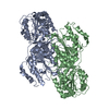
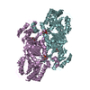
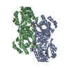
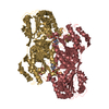
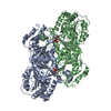
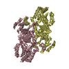
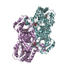
| Title | Crystal structure of menaquinone synthesis protein MenD from E. coli in complex with ThDP | ||||||
 Components Components | 2-succinyl-5-enolpyruvyl-6-hydroxy-3-cyclohexene-1-carboxylate synthase | ||||||
 Keywords Keywords |  TRANSFERASE / menaquinone / ThDP / Mg / vitamin K2 / carboxylase / Magnesium / Manganese / Menaquinone biosynthesis / Metal-binding / Thiamine pyrophosphate TRANSFERASE / menaquinone / ThDP / Mg / vitamin K2 / carboxylase / Magnesium / Manganese / Menaquinone biosynthesis / Metal-binding / Thiamine pyrophosphate | ||||||
| Function / homology |  Function and homology information2-succinyl-5-enolpyruvyl-6-hydroxy-3-cyclohexene-1-carboxylic-acid synthase / 2-succinyl-5-enolpyruvyl-6-hydroxy-3-cyclohexene-1-carboxylic-acid synthase activity / menaquinone biosynthetic process / thiamine pyrophosphate binding / manganese ion binding / magnesium ion binding / protein homodimerization activity Function and homology information2-succinyl-5-enolpyruvyl-6-hydroxy-3-cyclohexene-1-carboxylic-acid synthase / 2-succinyl-5-enolpyruvyl-6-hydroxy-3-cyclohexene-1-carboxylic-acid synthase activity / menaquinone biosynthetic process / thiamine pyrophosphate binding / manganese ion binding / magnesium ion binding / protein homodimerization activitySimilarity search - Function | ||||||
| Biological species |  Escherichia coli (E. coli) Escherichia coli (E. coli) | ||||||
| Method | X-RAY DIFFRACTION / SYNCHROTRON / MOLECULAR REPLACEMENT / Resolution: 2.6 Å | ||||||
 Authors Authors | Priyadarshi, A. / Hwang, K.Y. | ||||||
 Citation Citation | Journal: Biochem.Biophys.Res.Commun. / Year: 2009 Title: Structural and functional analysis of Vitamin K2 synthesis protein MenD. Authors: Priyadarshi, A. / Kim, E.E. / Hwang, K.Y. | ||||||
| History |
|
- Structure visualization
| Structure viewer | Molecule: MolmilJmol/JSmol |
|---|
- Downloads & links
-Download
| PDBx/mmCIF format | 3hwx.cif.gz | 841.2 KB | Display | PDBx/mmCIF format |
|---|---|---|---|---|
| PDB format | pdb3hwx.ent.gz | 693.8 KB | Display | PDB format |
| PDBx/mmJSON format | 3hwx.json.gz | Tree view | PDBx/mmJSON format | |
| Others |  Other downloads Other downloads |
-Validation report
| Arichive directory | https://data.pdbj.org/pub/pdb/validation_reports/hw/3hwxftp://data.pdbj.org/pub/pdb/validation_reports/hw/3hwx | HTTPS FTP |
|---|
-Related structure data
| Related structure data |  3hwwC  2jlcS S: Starting model for refinement C: citing same article ( |
|---|---|
| Similar structure data |


-Links
 PDBj
PDBj


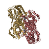
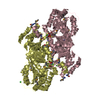
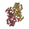
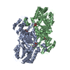
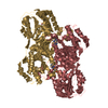
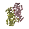
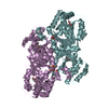
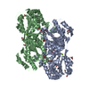
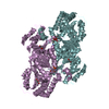
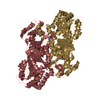
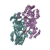
- Assembly
| Deposited unit | 
| ||||||||
|---|---|---|---|---|---|---|---|---|---|
| 1 | 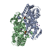
| ||||||||
| 2 | 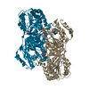
| ||||||||
| 3 | 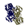
| ||||||||
| 4 | 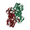
| ||||||||
| 5 | 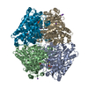
| ||||||||
| 6 | 
| ||||||||
| Unit cell |
|
-Components
-Protein , 1 types, 8 molecules ABIJRSZ1
| #1: Protein | Mass: 61449.828 Da / Num. of mol.: 8 / Mutation: P36L Source method: isolated from a genetically manipulated source Source: (gene. exp.) Escherichia coli (E. coli) / Strain: K12 / Gene: b2264, JW5374, menD / Plasmid: pET28a / Production host: Escherichia coli (E. coli) / Strain (production host): BL21 (DE3)References: UniProt: P17109, 2-succinyl-5-enolpyruvyl-6-hydroxy-3-cyclohexene-1-carboxylic-acid synthase |
|---|
-Non-polymers , 5 types, 484 molecules 


| #2: Chemical | ChemComp-NA /  Mass: 22.990 Da / Num. of mol.: 15 / Source method: obtained synthetically / Formula: Na Mass: 22.990 Da / Num. of mol.: 15 / Source method: obtained synthetically / Formula: Na#3: Chemical | ChemComp-TPP / Thiamine pyrophosphate Mass: 425.314 Da / Num. of mol.: 8 / Source method: obtained synthetically / Formula: C12H19N4O7P2S Mass: 425.314 Da / Num. of mol.: 8 / Source method: obtained synthetically / Formula: C12H19N4O7P2S#4: Chemical | ChemComp-MG /  Mass: 24.305 Da / Num. of mol.: 8 / Source method: obtained synthetically / Formula: Mg Mass: 24.305 Da / Num. of mol.: 8 / Source method: obtained synthetically / Formula: Mg#5: Chemical | Glycerol Mass: 92.094 Da / Num. of mol.: 2 / Source method: obtained synthetically / Formula: C3H8O3 Mass: 92.094 Da / Num. of mol.: 2 / Source method: obtained synthetically / Formula: C3H8O3#6: Water | ChemComp-HOH / | WaterMass: 18.015 Da / Num. of mol.: 451 / Source method: isolated from a natural source / Formula: H2O |
|---|
-Experimental details
-Experiment
| Experiment | Method: X-RAY DIFFRACTION / Number of used crystals: 1 |
|---|
- Sample preparation
| Crystal | Density Matthews: 2.46 Å3/Da / Density % sol: 49.91 % |
|---|---|
| Crystal grow | Temperature: 295 K / Method: vapor diffusion, hanging drop / pH: 7.5 Details: 12% PEG 8K, 10% glycerol, 1mM ThDP, 5mM MgCl2, pH 7.5, VAPOR DIFFUSION, HANGING DROP, temperature 295K |
-Data collection
| Diffraction | Mean temperature: 100 K |
|---|---|
| Diffraction source | Source: SYNCHROTRON / Site: PAL/PLS  / Beamline: 6C1 / Wavelength: 1 Å / Beamline: 6C1 / Wavelength: 1 Å |
| Detector | Type: ADSC QUANTUM 270 / Detector: CCD / Date: May 13, 2009 / Details: mirrors |
| Radiation | Monochromator: GRAPHITE / Protocol: SINGLE WAVELENGTH / Monochromatic (M) / Laue (L): M / Scattering type: x-ray |
| Radiation wavelength | Wavelength: 1 Å / Relative weight: 1 |
| Reflection | Resolution: 2.4→50 Å / Num. all: 141599 / Num. obs: 114098 / % possible obs: 83.5 % / Observed criterion σ(F): 0 / Observed criterion σ(I): 0 / Redundancy: 2.4 % / Biso Wilson estimate: 14.5 Å2 / Rmerge(I) obs: 0.15 / Rsym value: 0.26 / Net I/σ(I): 6.22 |
| Reflection shell | Resolution: 2.4→2.44 Å / Redundancy: 1.5 % / Rmerge(I) obs: 0.15 / Mean I/σ(I) obs: 1.8 / Num. unique all: 4413 / Rsym value: 0.26 / % possible all: 48.1 |
- Processing
| Software |
| |||||||||||||||||||||||||||||||||||||||||||||||||||||||||||||||||
|---|---|---|---|---|---|---|---|---|---|---|---|---|---|---|---|---|---|---|---|---|---|---|---|---|---|---|---|---|---|---|---|---|---|---|---|---|---|---|---|---|---|---|---|---|---|---|---|---|---|---|---|---|---|---|---|---|---|---|---|---|---|---|---|---|---|---|
| Refinement | Method to determine structure: MOLECULAR REPLACEMENT Starting model: 2JLC Resolution: 2.6→47.69 Å / Cor.coef. Fo:Fc: 0.898 / Cor.coef. Fo:Fc free: 0.835 / SU B: 11.452 / SU ML: 0.249 / Cross valid method: THROUGHOUT / σ(F): 0 / σ(I): 0 / ESU R Free: 0.395 / Stereochemistry target values: MAXIMUM LIKELIHOOD / Details: HYDROGENS HAVE BEEN ADDED IN THE RIDING POSITIONS
| |||||||||||||||||||||||||||||||||||||||||||||||||||||||||||||||||
| Solvent computation | Ion probe radii: 0.8 Å / Shrinkage radii: 0.8 Å / VDW probe radii: 1.4 Å / Solvent model: MASK | |||||||||||||||||||||||||||||||||||||||||||||||||||||||||||||||||
| Displacement parameters | Biso mean: 14.503 Å2
| |||||||||||||||||||||||||||||||||||||||||||||||||||||||||||||||||
| Refinement step | Cycle: LAST / Resolution: 2.6→47.69 Å
| |||||||||||||||||||||||||||||||||||||||||||||||||||||||||||||||||
| Refine LS restraints |
| |||||||||||||||||||||||||||||||||||||||||||||||||||||||||||||||||
| LS refinement shell | Resolution: 2.6→2.667 Å / Total num. of bins used: 20
|
