 Movie
Movie Controller

[English] 日本語
 Yorodumi
- PDB-3hsw: Crystal Structure of Porcine Pancreatic Phospholipase A2 in Compl... -
+ Open data

- Basic information
| Entry | Database: PDB / ID: 3hsw | ||||||
|---|---|---|---|---|---|---|---|
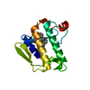
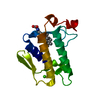
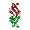
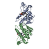
| Title | Crystal Structure of Porcine Pancreatic Phospholipase A2 in Complex with 2-methoxycyclohexa-2-5-diene-1,4-dione | ||||||
 Components Components | Phospholipase A2, major isoenzyme | ||||||
 Keywords Keywords | HYDROLASE / Curcumin binding / PLA2 / Pancreatic Enzyme / Disulfide bond / Lipid degradation / Lipoprotein / Metal-binding / Palmitate / Pyrrolidone carboxylic acid / Secreted | ||||||
| Function / homology |  Function and homology information Function and homology informationAcyl chain remodelling of PC / Acyl chain remodelling of PS / Acyl chain remodelling of PE / Acyl chain remodelling of PI / Acyl chain remodelling of PG / Synthesis of PA / positive regulation of podocyte apoptotic process / regulation of glucose import / phosphatidylglycerol metabolic process / phosphatidylcholine metabolic process ...Acyl chain remodelling of PC / Acyl chain remodelling of PS / Acyl chain remodelling of PE / Acyl chain remodelling of PI / Acyl chain remodelling of PG / Synthesis of PA / positive regulation of podocyte apoptotic process / regulation of glucose import / phosphatidylglycerol metabolic process / phosphatidylcholine metabolic process / phospholipase A2 activity / leukotriene biosynthetic process / neutrophil mediated immunity / calcium-dependent phospholipase A2 activity / phospholipase A2 / bile acid binding / positive regulation of calcium ion transport into cytosol / phospholipid metabolic process / lipid catabolic process / neutrophil chemotaxis / positive regulation of interleukin-8 production / positive regulation of MAP kinase activity / phospholipid binding / fatty acid biosynthetic process / cellular response to insulin stimulus / positive regulation of immune response / positive regulation of fibroblast proliferation / positive regulation of NF-kappaB transcription factor activity / intracellular signal transduction / signaling receptor binding / calcium ion binding / positive regulation of cell population proliferation / cell surface / positive regulation of transcription by RNA polymerase II / extracellular regionSimilarity search - Function | ||||||
| Biological species |  Sus scrofa (pig) Sus scrofa (pig) | ||||||
| Method | X-RAY DIFFRACTION / MOLECULAR REPLACEMENT / Resolution: 2.5 Å | ||||||
 Authors Authors | Dileep, K.V. / Tintu, I. / Karthe, P. / Mandal, P.K. / Haridas, M. / Sadasivan, C. | ||||||
 Citation Citation | Journal: Frontiers in life sci. / Year: 2012 Title: Crystal structure of porcine pancreatic phospholipase A2 in complex with 2-methoxycyclohexa-2-5-diene-1,4-dione Authors: Dileep, K.V. / Tintu, I. / Mandal, P.K. / Karthe, P. / Haridas, M. / Sadasivan, C. | ||||||
| History |
|
- Structure visualization
| Structure viewer | Molecule: MolmilJmol/JSmol |
|---|
- Downloads & links
-Download
| PDBx/mmCIF format | 3hsw.cif.gz | 39.1 KB | Display | PDBx/mmCIF format |
|---|---|---|---|---|
| PDB format | pdb3hsw.ent.gz | 26.4 KB | Display | PDB format |
| PDBx/mmJSON format | 3hsw.json.gz | Tree view | PDBx/mmJSON format | |
| Others |  Other downloads Other downloads |
-Validation report
| Arichive directory | https://data.pdbj.org/pub/pdb/validation_reports/hs/3hswftp://data.pdbj.org/pub/pdb/validation_reports/hs/3hsw | HTTPS FTP |
|---|
-Related structure data
| Related structure data | 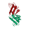 1p2pS S: Starting model for refinement |
|---|---|
| Similar structure data |


-Links
 PDBj
PDBj

- Assembly
| Deposited unit | 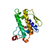
| ||||||||
|---|---|---|---|---|---|---|---|---|---|
| 1 | 
| ||||||||
| Unit cell |
| ||||||||
| Components on special symmetry positions |
|
-Components
| #1: Protein | / Phosphatidylcholine 2-acylhydrolase / Group IB phospholipase A2 Mass: 14009.714 Da / Num. of mol.: 1 / Source method: isolated from a natural source / Details: Pancreas / Source: (natural) Sus scrofa (pig) / References: UniProt: P00592, phospholipase A2 | ||
|---|---|---|---|
| #2: Chemical | ChemComp-MCW /   Mass: 138.121 Da / Num. of mol.: 1 / Source method: obtained synthetically / Formula: C7H6O3 Mass: 138.121 Da / Num. of mol.: 1 / Source method: obtained synthetically / Formula: C7H6O3 | ||
| #3: Chemical |   Mass: 40.078 Da / Num. of mol.: 2 / Source method: obtained synthetically / Formula: Ca Mass: 40.078 Da / Num. of mol.: 2 / Source method: obtained synthetically / Formula: Ca#4: Water | ChemComp-HOH / | Water Mass: 18.015 Da / Num. of mol.: 32 / Source method: isolated from a natural source / Formula: H2O Mass: 18.015 Da / Num. of mol.: 32 / Source method: isolated from a natural source / Formula: H2O |
-Experimental details
-Experiment
| Experiment | Method: X-RAY DIFFRACTION / Number of used crystals: 1 |
|---|
- Sample preparation
| Crystal | Density Matthews: 3.38 Å3/Da / Density % sol: 63.57 % |
|---|---|
| Crystal grow | Temperature: 294 K / Method: vapor diffusion, hanging drop / pH: 7.2 Details: 15% MPD, 0.05M Tris Maleate, 5mM Calcium Chloride, pH7.2, VAPOR DIFFUSION, HANGING DROP, temperature 294K |
-Data collection
| Diffraction | Mean temperature: 100 K |
|---|---|
| Diffraction source | Source: ROTATING ANODE / Type: BRUKER AXS MICROSTAR / Wavelength: 1.542 Å |
| Detector | Type: MAR scanner 345 mm plate / Detector: IMAGE PLATE / Date: Mar 22, 2009 / Details: Mirrors |
| Radiation | Protocol: SINGLE WAVELENGTH / Monochromatic (M) / Laue (L): M / Scattering type: x-ray |
| Radiation wavelength | Wavelength: 1.542 Å / Relative weight: 1 |
| Reflection | Resolution: 2.5→45.24 Å / Num. all: 6789 / Num. obs: 6759 / % possible obs: 97.3 % / Observed criterion σ(F): 2.5 / Observed criterion σ(I): 2 / Rmerge(I) obs: 0.073 / Rsym value: 0.065 / Net I/σ(I): 8.5 |
| Reflection shell | Resolution: 2.5→2.64 Å / Rmerge(I) obs: 0.073 / Num. unique all: 955 / Rsym value: 0.065 / % possible all: 70.79 |
- Processing
| Software |
| |||||||||||||||||||||||||||||||||||||||||||||||||||||||||||||||||
|---|---|---|---|---|---|---|---|---|---|---|---|---|---|---|---|---|---|---|---|---|---|---|---|---|---|---|---|---|---|---|---|---|---|---|---|---|---|---|---|---|---|---|---|---|---|---|---|---|---|---|---|---|---|---|---|---|---|---|---|---|---|---|---|---|---|---|
| Refinement | Method to determine structure: MOLECULAR REPLACEMENT Starting model: PDB ENTRY 1P2P Resolution: 2.5→13.65 Å / Cor.coef. Fo:Fc: 0.952 / Cor.coef. Fo:Fc free: 0.923 / SU B: 17.329 / SU ML: 0.174 / TLS residual ADP flag: LIKELY RESIDUAL / Isotropic thermal model: ISOTROPIC / Cross valid method: THROUGHOUT / σ(F): 2.5 / σ(I): 2 / ESU R: 0.333 / ESU R Free: 0.241 / Stereochemistry target values: MAXIMUM LIKELIHOOD / Details: HYDROGENS HAVE BEEN ADDED IN THE RIDING POSITIONS
| |||||||||||||||||||||||||||||||||||||||||||||||||||||||||||||||||
| Solvent computation | Ion probe radii: 0.8 Å / Shrinkage radii: 0.8 Å / VDW probe radii: 1.4 Å / Solvent model: MASK | |||||||||||||||||||||||||||||||||||||||||||||||||||||||||||||||||
| Displacement parameters | Biso mean: 23.783 Å2
| |||||||||||||||||||||||||||||||||||||||||||||||||||||||||||||||||
| Refinement step | Cycle: LAST / Resolution: 2.5→13.65 Å
| |||||||||||||||||||||||||||||||||||||||||||||||||||||||||||||||||
| Refine LS restraints |
| |||||||||||||||||||||||||||||||||||||||||||||||||||||||||||||||||
| LS refinement shell | Resolution: 2.5→2.563 Å / Total num. of bins used: 20
| |||||||||||||||||||||||||||||||||||||||||||||||||||||||||||||||||
| Refinement TLS params. | Method: refined / Origin x: -35.291 Å / Origin y: 14.13 Å / Origin z: -2.717 Å
|
