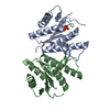
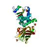
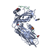
[English] 日本語
Yorodumi
- PDB-3hs8: Intersectin 1-peptide-AP2 alpha ear complex -

+
Open data


ID or keywords:

Loading...

-
Basic information

Entry
Database: PDB / ID: 3hs8
TitleIntersectin 1-peptide-AP2 alpha ear complex
Components
  • Adaptor protein complex AP-2, alpha 2 subunitAdapter
  • peptide from Intersectin-1, residues 840-851
KeywordsENDOCYTOSIS / adaptor complex AP-2 / Cell membrane / Coated pit / Lipid-binding / Membrane / Disease mutation / Transferase
Function / homology
Function and homology information


presynaptic endocytic zone / RHOQ GTPase cycle / positive regulation of caveolin-mediated endocytosis / clathrin-dependent synaptic vesicle endocytosis / CDC42 GTPase cycle / EPHB-mediated forward signaling / NRAGE signals death through JNK / LDL clearance / WNT5A-dependent internalization of FZD2, FZD5 and ROR2 / WNT5A-dependent internalization of FZD4 ...presynaptic endocytic zone / RHOQ GTPase cycle / positive regulation of caveolin-mediated endocytosis / clathrin-dependent synaptic vesicle endocytosis / CDC42 GTPase cycle / EPHB-mediated forward signaling / NRAGE signals death through JNK / LDL clearance / WNT5A-dependent internalization of FZD2, FZD5 and ROR2 / WNT5A-dependent internalization of FZD4 / Trafficking of GluR2-containing AMPA receptors / Retrograde neurotrophin signalling / RHOG GTPase cycle / VLDLR internalisation and degradation / G alpha (12/13) signalling events / Recycling pathway of L1 / AP-2 adaptor complex / positive regulation of growth hormone secretion / postsynaptic neurotransmitter receptor internalization / Cargo recognition for clathrin-mediated endocytosis / postsynaptic actin cytoskeleton / clathrin adaptor activity / Clathrin-mediated endocytosis / regulation of modification of postsynaptic actin cytoskeleton / membrane coat / clathrin-dependent endocytosis / apical dendrite / MHC class II antigen presentation / kinase activator activity / regulation of hematopoietic stem cell differentiation / regulation of postsynapse organization / endosomal transport / small GTPase-mediated signal transduction / positive regulation of Rho protein signal transduction / protein serine/threonine kinase binding / intracellular vesicle / positive regulation of dendritic spine development / exocytosis / calyx of Held / synaptic vesicle endocytosis / endocytic vesicle / clathrin-coated pit / vesicle-mediated transport / Neutrophil degranulation / phosphatidylinositol binding / guanyl-nucleotide exchange factor activity / secretory granule / intracellular protein transport / recycling endosome / cytoplasmic side of plasma membrane / kinase binding / disordered domain specific binding / synaptic vesicle / protein transport / lamellipodium / presynaptic membrane / nuclear envelope / cytoplasmic vesicle / postsynapse / neuron apoptotic process / negative regulation of neuron apoptotic process / dendritic spine / positive regulation of phosphatidylinositol 3-kinase/protein kinase B signal transduction / molecular adaptor activity / protein domain specific binding / neuronal cell body / glutamatergic synapse / calcium ion binding / protein-containing complex binding / protein kinase binding / plasma membrane / cytosol / cytoplasm
Similarity search - Function
Intersectin-1, AP2 binding region / Intersectin and clathrin adaptor AP2 binding region / Pleckstrin homology domain / Gamma-adaptin ear (GAE) domain / Clathrin adaptor, alpha-adaptin, appendage, C-terminal subdomain / Adaptor protein complex AP-2, alpha subunit / Alpha adaptin AP2, C-terminal domain / Cytoskeletal-regulatory complex EF hand / EH domain profile. / Eps15 homology domain ...Intersectin-1, AP2 binding region / Intersectin and clathrin adaptor AP2 binding region / Pleckstrin homology domain / Gamma-adaptin ear (GAE) domain / Clathrin adaptor, alpha-adaptin, appendage, C-terminal subdomain / Adaptor protein complex AP-2, alpha subunit / Alpha adaptin AP2, C-terminal domain / Cytoskeletal-regulatory complex EF hand / EH domain profile. / Eps15 homology domain / EH domain / TATA-Binding Protein / Guanine-nucleotide dissociation stimulator, CDC24, conserved site / Dbl homology (DH) domain signature. / Coatomer/calthrin adaptor appendage, C-terminal subdomain / Clathrin adaptor, alpha/beta/gamma-adaptin, appendage, Ig-like subdomain / Adaptin C-terminal domain / Adaptin C-terminal domain / Clathrin/coatomer adaptor, adaptin-like, N-terminal / Adaptin N terminal region / Clathrin adaptor, appendage, Ig-like subdomain superfamily / Variant SH3 domain / TATA-Binding Protein / Variant SH3 domain / Dbl homology (DH) domain superfamily / RhoGEF domain / Guanine nucleotide exchange factor for Rho/Rac/Cdc42-like GTPases / Dbl homology (DH) domain / Dbl homology (DH) domain profile. / TBP domain superfamily / C2 domain / Protein kinase C conserved region 2 (CalB) / C2 domain / C2 domain profile. / C2 domain superfamily / PH domain profile. / Pleckstrin homology domain. / Pleckstrin homology domain / SH3 domain / EF-hand, calcium binding motif / Src homology 3 domains / EF-Hand 1, calcium-binding site / EF-hand calcium-binding domain. / EF-hand calcium-binding domain profile. / SH3-like domain superfamily / Src homology 3 (SH3) domain profile. / SH3 domain / EF-hand domain / Armadillo-like helical / EF-hand domain pair / PH-like domain superfamily / Armadillo-type fold / Immunoglobulin-like / Sandwich / 2-Layer Sandwich / Mainly Beta / Alpha Beta
Similarity search - Domain/homology
AP-2 complex subunit alpha-2 / AP-2 complex subunit alpha / Intersectin-1
Similarity search - Component
Biological speciesMus musculus (house mouse)
MethodX-RAY DIFFRACTION / SYNCHROTRON / MOLECULAR REPLACEMENT / molecular replacement / Resolution: 1.9 Å
AuthorsVahedi-Faridi, A. / Pechstein, A. / Schaefer, J.G. / Saenger, W. / Haucke, V.
CitationJournal: Proc.Natl.Acad.Sci.USA / Year: 2010
Title: Regulation of synaptic vesicle recycling by complex formation between intersectin 1 and the clathrin adaptor complex AP2.
Authors: Pechstein, A. / Bacetic, J. / Vahedi-Faridi, A. / Gromova, K. / Sundborger, A. / Tomlin, N. / Krainer, G. / Vorontsova, O. / Schafer, J.G. / Owe, S.G. / Cousin, M.A. / Saenger, W. / Shupliakov, O. / Haucke, V.
History
DepositionJun 10, 2009Deposition site: RCSB / Processing site: PDBJ
Revision 1.0Feb 23, 2010Provider: repository / Type: Initial release
Revision 1.1Jul 13, 2011Group: Advisory / Refinement description / Version format compliance
Revision 1.2Nov 1, 2017Group: Refinement description / Category: software
Revision 1.3Mar 20, 2024Group: Data collection / Database references
Category: chem_comp_atom / chem_comp_bond ...chem_comp_atom / chem_comp_bond / database_2 / struct_ref_seq_dif
Item: _database_2.pdbx_DOI / _database_2.pdbx_database_accession / _struct_ref_seq_dif.details

-
Structure visualization

Structure viewerMolecule:
MolmilJmol/JSmol

Downloads & links

-
Assembly

Deposited unit
A: Adaptor protein complex AP-2, alpha 2 subunit
P: peptide from Intersectin-1, residues 840-851


Theoretical massNumber of molelcules
Total (without water)32,1662
Polymers32,1662
Non-polymers00
Water2,882160
1


  • Idetical with deposited unit
  • defined by author&software
TypeNameSymmetry operationNumber
identity operation1_555x,y,z1
Buried area790 Å2
ΔGint-4 kcal/mol
Surface area13070 Å2
MethodPISA
Unit cell
Length a, b, c (Å)64.484, 120.412, 32.220
Angle α, β, γ (deg.)90.000, 90.000, 90.000
Int Tables number18
Space group name H-MP21212

-
Components

#1: Protein Adaptor protein complex AP-2, alpha 2 subunit / Adapter


Mass: 30744.889 Da / Num. of mol.: 1 / Fragment: UNP residues 702-938
Source method: isolated from a genetically manipulated source
Source: (gene. exp.) Mus musculus (house mouse) / Gene: Ap2a2 / Plasmid: pET-28(+) / Production host: Escherichia coli (E. coli) / Strain (production host): BL21(DE3) / References: UniProt: Q6PEE6, UniProt: P17427*PLUS
#2: Protein/peptide peptide from Intersectin-1, residues 840-851 /


Mass: 1421.469 Da / Num. of mol.: 1 / Source method: obtained synthetically / Details: synthetic peptide / References: UniProt: Q9Z0R4
#3: Water ChemComp-HOH / water / Water


Mass: 18.015 Da / Num. of mol.: 160 / Source method: isolated from a natural source / Formula: H2O

-
Experimental details

-
Experiment

ExperimentMethod: X-RAY DIFFRACTION / Number of used crystals: 1

-
Sample preparation

CrystalDensity Matthews: 1.94 Å3/Da / Density % sol: 36.74 % / Mosaicity: 0.709 °
Crystal growTemperature: 291 K / Method: vapor diffusion, hanging drop / pH: 8
Details: 20% PEG4000, 100mM HEPES, 10% Isopropanol, pH 8.0, VAPOR DIFFUSION, HANGING DROP, temperature 291.0K

-
Data collection

DiffractionMean temperature: 100 K
Diffraction sourceSource: SYNCHROTRON / Site: BESSY / Beamline: 14.2 / Wavelength: 0.91841 Å
DetectorType: MARMOSAIC 225 mm CCD / Detector: CCD / Date: Jul 17, 2007 / Details: mirrors
RadiationMonochromator: Si 111 CHANNEL / Protocol: SINGLE WAVELENGTH / Monochromatic (M) / Laue (L): M / Scattering type: x-ray
Radiation wavelengthWavelength: 0.91841 Å / Relative weight: 1
ReflectionResolution: 1.9→40 Å / Num. obs: 19195 / % possible obs: 93.3 % / Observed criterion σ(F): 2 / Redundancy: 2.9 % / Rmerge(I) obs: 0.03 / Χ2: 0.733 / Net I/σ(I): 27.513
Reflection shell
Resolution (Å)Redundancy (%)Rmerge(I) obsNum. unique allΧ2Diffraction-ID% possible all
1.9-1.972.50.1316300.838183.2
1.97-2.052.80.11318760.815192.7
2.05-2.142.90.07918370.748191.2
2.14-2.252.90.06318720.736192.3
2.25-2.3930.05218880.721192.4
2.39-2.5830.04218950.704192.6
2.58-2.8430.03319570.677195.3
2.84-3.2530.02720470.741198.6
3.25-4.0930.02820790.565199
4.09-402.90.01921140.836194.4

-
Phasing

PhasingMethod: molecular replacement
Phasing MR
Highest resolutionLowest resolution
Rotation2.05 Å31.12 Å
Translation2.05 Å31.12 Å

-
Processing

Software
NameVersionClassificationNB
DENZOdata reduction
SCALEPACKdata scaling
MOLREPphasing
REFMACrefinement
PDB_EXTRACT3.005data extraction
RefinementMethod to determine structure: MOLECULAR REPLACEMENT / Resolution: 1.9→30.1 Å / Cor.coef. Fo:Fc: 0.952 / Cor.coef. Fo:Fc free: 0.927 / WRfactor Rfree: 0.251 / WRfactor Rwork: 0.226 / Occupancy max: 1 / Occupancy min: 0.5 / FOM work R set: 0.845 / SU B: 7.717 / SU ML: 0.104 / SU R Cruickshank DPI: 0.208 / SU Rfree: 0.173 / TLS residual ADP flag: LIKELY RESIDUAL / Cross valid method: THROUGHOUT / σ(F): 0 / ESU R: 0.178 / ESU R Free: 0.161 / Stereochemistry target values: MAXIMUM LIKELIHOOD / Details: HYDROGENS HAVE BEEN ADDED IN THE RIDING POSITIONS
RfactorNum. reflection% reflectionSelection details
Rfree0.235 1000 5.2 %RANDOM
Rwork0.189 ---
obs0.191 19174 93.39 %-
Solvent computationIon probe radii: 0.8 Å / Shrinkage radii: 0.8 Å / VDW probe radii: 1.2 Å / Solvent model: MASK
Displacement parametersBiso max: 52.96 Å2 / Biso mean: 27.451 Å2 / Biso min: 12.55 Å2
Baniso -1Baniso -2Baniso -3
1--0.43 Å20 Å20 Å2
2--0.11 Å20 Å2
3---0.32 Å2
Refinement stepCycle: LAST / Resolution: 1.9→30.1 Å
ProteinNucleic acidLigandSolventTotal
Num. atoms2005 0 0 160 2165
Refine LS restraints
Refine-IDTypeDev idealDev ideal targetNumber
X-RAY DIFFRACTIONr_bond_refined_d0.0180.0222049
X-RAY DIFFRACTIONr_angle_refined_deg1.6451.9442779
X-RAY DIFFRACTIONr_dihedral_angle_1_deg7.3865250
X-RAY DIFFRACTIONr_dihedral_angle_2_deg38.79425.619105
X-RAY DIFFRACTIONr_dihedral_angle_3_deg15.35415354
X-RAY DIFFRACTIONr_dihedral_angle_4_deg21.792159
X-RAY DIFFRACTIONr_chiral_restr0.1350.2311
X-RAY DIFFRACTIONr_gen_planes_refined0.0070.021571
X-RAY DIFFRACTIONr_nbd_refined0.2110.2891
X-RAY DIFFRACTIONr_nbtor_refined0.3080.21389
X-RAY DIFFRACTIONr_xyhbond_nbd_refined0.1530.2148
X-RAY DIFFRACTIONr_symmetry_vdw_refined0.2540.261
X-RAY DIFFRACTIONr_symmetry_hbond_refined0.2390.212
X-RAY DIFFRACTIONr_mcbond_it0.9221.51299
X-RAY DIFFRACTIONr_mcangle_it1.3922037
X-RAY DIFFRACTIONr_scbond_it2.4743851
X-RAY DIFFRACTIONr_scangle_it3.8854.5741
LS refinement shellResolution: 1.899→1.949 Å / Total num. of bins used: 20
RfactorNum. reflection% reflection
Rfree0.251 87 -
Rwork0.21 1083 -
all-1170 -
obs--81.02 %
Refinement TLS params.

Method: refined / Refine-ID: X-RAY DIFFRACTION

IDL112)L122)L132)L222)L232)L332)S11 (Å °)S12 (Å °)S13 (Å °)S21 (Å °)S22 (Å °)S23 (Å °)S31 (Å °)S32 (Å °)S33 (Å °)T112)T122)T132)T222)T232)T332)Origin x (Å)Origin y (Å)Origin z (Å)
12.1101-0.42340.10050.92470.30362.23450.0910.0176-0.2719-0.0229-0.0750.00280.35510.0925-0.0159-0.11430.017-0.0158-0.12870.018-0.094941.116417.894112.6087
210.48912.004110.718513.922910.429729.20260.31441.1830.3015-1.03470.4809-0.7368-0.73033.2231-0.79530.16980.10330.02250.75650.0090.436659.396513.534-0.5678
31.5277-0.15360.1730.80710.64811.85650.05510.0105-0.0595-0.0367-0.0133-0.04080.06820.0584-0.0418-0.0121-0.00930.0053-0.03040.01820.015640.236223.678315.0244
Refinement TLS group
IDRefine-IDRefine TLS-IDAuth asym-IDAuth seq-ID
1X-RAY DIFFRACTION1A701 - 810
2X-RAY DIFFRACTION1A811 - 938
3X-RAY DIFFRACTION2P840 - 851
4X-RAY DIFFRACTION3A1 - 120

+
About Yorodumi

-
News

-
Feb 9, 2022. New format data for meta-information of EMDB entries

New format data for meta-information of EMDB entries

  • Version 3 of the EMDB header file is now the official format.
  • The previous official version 1.9 will be removed from the archive.

Related info.:EMDB header

External links:wwPDB to switch to version 3 of the EMDB data model

-
Aug 12, 2020. Covid-19 info

Covid-19 info

URL: https://pdbj.org/emnavi/covid19.php

New page: Covid-19 featured information page in EM Navigator.

Related info.:Covid-19 info / Mar 5, 2020. Novel coronavirus structure data

+
Mar 5, 2020. Novel coronavirus structure data

Novel coronavirus structure data

Related info.:Yorodumi Speices / Aug 12, 2020. Covid-19 info

External links:COVID-19 featured content - PDBj / Molecule of the Month (242):Coronavirus Proteases

+
Jan 31, 2019. EMDB accession codes are about to change! (news from PDBe EMDB page)

EMDB accession codes are about to change! (news from PDBe EMDB page)

  • The allocation of 4 digits for EMDB accession codes will soon come to an end. Whilst these codes will remain in use, new EMDB accession codes will include an additional digit and will expand incrementally as the available range of codes is exhausted. The current 4-digit format prefixed with “EMD-” (i.e. EMD-XXXX) will advance to a 5-digit format (i.e. EMD-XXXXX), and so on. It is currently estimated that the 4-digit codes will be depleted around Spring 2019, at which point the 5-digit format will come into force.
  • The EM Navigator/Yorodumi systems omit the EMD- prefix.

Related info.:Q: What is EMD? / ID/Accession-code notation in Yorodumi/EM Navigator

External links:EMDB Accession Codes are Changing Soon! / Contact to PDBj

+
Jul 12, 2017. Major update of PDB

Major update of PDB

  • wwPDB released updated PDB data conforming to the new PDBx/mmCIF dictionary.
  • This is a major update changing the version number from 4 to 5, and with Remediation, in which all the entries are updated.
  • In this update, many items about electron microscopy experimental information are reorganized (e.g. em_software).
  • Now, EM Navigator and Yorodumi are based on the updated data.

External links:wwPDB Remediation / Enriched Model Files Conforming to OneDep Data Standards Now Available in the PDB FTP Archive

-
Yorodumi

Thousand views of thousand structures

  • Yorodumi is a browser for structure data from EMDB, PDB, SASBDB, etc.
  • This page is also the successor to EM Navigator detail page, and also detail information page/front-end page for Omokage search.
  • The word "yorodu" (or yorozu) is an old Japanese word meaning "ten thousand". "mi" (miru) is to see.

Related info.:EMDB / PDB / SASBDB / Comparison of 3 databanks / Yorodumi Search / Aug 31, 2016. New EM Navigator & Yorodumi / Yorodumi Papers / Jmol/JSmol / Function and homology information / Changes in new EM Navigator and Yorodumi

Read more