 Movie
Movie Controller


+ Open data

- Basic information
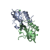
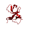
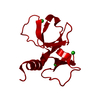
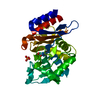
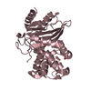
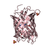
| Entry | Database: PDB / ID: 3hpw | ||||||
|---|---|---|---|---|---|---|---|
| Title | CcdB dimer in complex with one C-terminal CcdA domain | ||||||
 Components Components |
| ||||||
 Keywords Keywords | TOXIN/TOXIN REPRESSOR / ALPHA+BETA /  SH3 domain / intrinsically disordered / TOXIN-TOXIN REPRESSOR COMPLEX SH3 domain / intrinsically disordered / TOXIN-TOXIN REPRESSOR COMPLEX | ||||||
| Function / homology |  Function and homology information Function and homology informationDNA topoisomerase type II (double strand cut, ATP-hydrolyzing) inhibitor activity / toxin sequestering activity / negative regulation of DNA-templated DNA replication / protein-containing complex disassembly / toxin-antitoxin complex / plasmid maintenance / transcription repressor complex / negative regulation of DNA-templated transcription / DNA bindingSimilarity search - Function | ||||||
| Biological species |  Escherichia coli (E. coli) Escherichia coli (E. coli) | ||||||
| Method | X-RAY DIFFRACTION / SYNCHROTRON / MOLECULAR REPLACEMENT / Resolution: 1.452 Å | ||||||
 Authors Authors | De Jonge, N. / Loris, R. / Garcia-Pino, A. / Buts, L. | ||||||
 Citation Citation | Journal: Mol.Cell / Year: 2009 Title: Rejuvenation of CcdB-Poisoned Gyrase by an Intrinsically Disordered Protein Domain. Authors: De Jonge, N. / Garcia-Pino, A. / Buts, L. / Haesaerts, S. / Charlier, D. / Zangger, K. / Wyns, L. / De Greve, H. / Loris, R. | ||||||
| History |
|
- Structure visualization
| Structure viewer | Molecule: MolmilJmol/JSmol |
|---|
- Downloads & links
-Download
| PDBx/mmCIF format | 3hpw.cif.gz | 120.4 KB | Display | PDBx/mmCIF format |
|---|---|---|---|---|
| PDB format | pdb3hpw.ent.gz | 93.2 KB | Display | PDB format |
| PDBx/mmJSON format | 3hpw.json.gz | Tree view | PDBx/mmJSON format | |
| Others |  Other downloads Other downloads |
-Validation report
| Arichive directory | https://data.pdbj.org/pub/pdb/validation_reports/hp/3hpwftp://data.pdbj.org/pub/pdb/validation_reports/hp/3hpw | HTTPS FTP |
|---|
-Related structure data
| Related structure data | 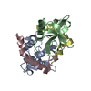 3g7zC 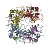 2vubS C: citing same article ( S: Starting model for refinement |
|---|---|
| Similar structure data |


-Links
 PDBj
PDBj- Assembly
| Deposited unit | 
| ||||||||
|---|---|---|---|---|---|---|---|---|---|
| 1 |
| ||||||||
| Unit cell |
|
-Components
| #1: Protein | Cytotoxicity / Protein letB / Protein G / LynB Mass: 11721.508 Da / Num. of mol.: 2 / Fragment: CcdB Source method: isolated from a genetically manipulated source Details: tac promotor / Source: (gene. exp.) Escherichia coli (E. coli) / Strain: JM101 / Gene: ccdB, ECOK12F043, G, letB / Plasmid: pKK223-3 / Production host: Escherichia coli (E. coli) / Strain (production host): CSH50 / References: UniProt: P62554#2: Protein/peptide | | Mass: 4376.829 Da / Num. of mol.: 1 / Fragment: C-terminal domain (UNP residues 37-72) / Source method: obtained synthetically Details: Fragment of the CcdA protein encoded on F-plasmid. Synthesised via solid phase synthesis by Bio-Synthesis. References: UniProt: P62552 #3: Chemical | ChemComp-PEG / | Diethylene glycol  Mass: 106.120 Da / Num. of mol.: 1 / Source method: obtained synthetically / Formula: C4H10O3 Mass: 106.120 Da / Num. of mol.: 1 / Source method: obtained synthetically / Formula: C4H10O3#4: Chemical | ChemComp-SO4 / | Sulfate  Mass: 96.063 Da / Num. of mol.: 1 / Source method: obtained synthetically / Formula: SO4 Mass: 96.063 Da / Num. of mol.: 1 / Source method: obtained synthetically / Formula: SO4#5: Water | ChemComp-HOH / | Water Mass: 18.015 Da / Num. of mol.: 329 / Source method: isolated from a natural source / Formula: H2O Mass: 18.015 Da / Num. of mol.: 329 / Source method: isolated from a natural source / Formula: H2O |
|---|
-Experimental details
-Experiment
| Experiment | Method: X-RAY DIFFRACTION / Number of used crystals: 1 |
|---|
- Sample preparation
| Crystal | Density Matthews: 1.95 Å3/Da / Density % sol: 37 % |
|---|---|
| Crystal grow | Temperature: 293 K / Method: vapor diffusion, hanging drop / pH: 7.4 Details: 22% PEG4000, 0.1M Tris-HCl pH7.4, 0.2M LiSO4, VAPOR DIFFUSION, HANGING DROP, temperature 293K |
-Data collection
| Diffraction | Mean temperature: 100 K |
|---|---|
| Diffraction source | Source: SYNCHROTRON / Site: EMBL/DESY, HAMBURG  / Beamline: BW7B / Wavelength: 0.8423 Å / Beamline: BW7B / Wavelength: 0.8423 Å |
| Detector | Type: MAR scanner 345 mm plate / Detector: IMAGE PLATE / Date: Jun 16, 2006 |
| Radiation | Protocol: SINGLE WAVELENGTH / Monochromatic (M) / Laue (L): M / Scattering type: x-ray |
| Radiation wavelength | Wavelength: 0.8423 Å / Relative weight: 1 |
| Reflection | Resolution: 1.45→15.78 Å / Num. all: 36688 / Num. obs: 36600 / % possible obs: 95.9 % / Observed criterion σ(F): 0 / Observed criterion σ(I): -3 / Redundancy: 2.9 % / Biso Wilson estimate: 13.4 Å2 / Rmerge(I) obs: 0.043 / Rsym value: 0.043 / Net I/σ(I): 18.3 |
| Reflection shell | Resolution: 1.45→1.5 Å / Redundancy: 2.9 % / Rmerge(I) obs: 0.107 / Mean I/σ(I) obs: 7.8 / Num. unique all: 3705 / Rsym value: 0.107 / % possible all: 98.2 |
- Processing
| Software |
| ||||||||||||||||||||||||||||||||||||||||||||||||||||||||||||||||||||||||||||||||||||||||||||||||||||
|---|---|---|---|---|---|---|---|---|---|---|---|---|---|---|---|---|---|---|---|---|---|---|---|---|---|---|---|---|---|---|---|---|---|---|---|---|---|---|---|---|---|---|---|---|---|---|---|---|---|---|---|---|---|---|---|---|---|---|---|---|---|---|---|---|---|---|---|---|---|---|---|---|---|---|---|---|---|---|---|---|---|---|---|---|---|---|---|---|---|---|---|---|---|---|---|---|---|---|---|---|---|
| Refinement | Method to determine structure: MOLECULAR REPLACEMENT Starting model: CcdB2 dimer, PDB entry 2VUB, chain A,B Resolution: 1.452→15.785 Å / Occupancy max: 1 / Occupancy min: 0.5 / FOM work R set: 0.892 / SU ML: 0.18 / Isotropic thermal model: anisotropic B factor refinement / Cross valid method: THROUGHOUT / σ(F): 1.36 / Phase error: 17.8 / Stereochemistry target values: ML
| ||||||||||||||||||||||||||||||||||||||||||||||||||||||||||||||||||||||||||||||||||||||||||||||||||||
| Solvent computation | Shrinkage radii: 0.9 Å / VDW probe radii: 1.11 Å / Solvent model: FLAT BULK SOLVENT MODEL / Bsol: 55.642 Å2 / ksol: 0.384 e/Å3 | ||||||||||||||||||||||||||||||||||||||||||||||||||||||||||||||||||||||||||||||||||||||||||||||||||||
| Displacement parameters | Biso max: 82.74 Å2 / Biso mean: 15.252 Å2 / Biso min: 4.34 Å2
| ||||||||||||||||||||||||||||||||||||||||||||||||||||||||||||||||||||||||||||||||||||||||||||||||||||
| Refinement step | Cycle: LAST / Resolution: 1.452→15.785 Å
| ||||||||||||||||||||||||||||||||||||||||||||||||||||||||||||||||||||||||||||||||||||||||||||||||||||
| Refine LS restraints |
| ||||||||||||||||||||||||||||||||||||||||||||||||||||||||||||||||||||||||||||||||||||||||||||||||||||
| LS refinement shell | Refine-ID: X-RAY DIFFRACTION / Total num. of bins used: 13
| ||||||||||||||||||||||||||||||||||||||||||||||||||||||||||||||||||||||||||||||||||||||||||||||||||||
| Refinement TLS params. | Method: refined / Refine-ID: X-RAY DIFFRACTION
| ||||||||||||||||||||||||||||||||||||||||||||||||||||||||||||||||||||||||||||||||||||||||||||||||||||
| Refinement TLS group |
|
