 Movie
Movie Controller


+ Open data

- Basic information
| Entry | Database: PDB / ID: 3hpb | ||||||
|---|---|---|---|---|---|---|---|
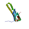
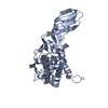
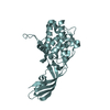
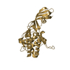
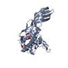
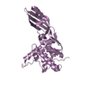
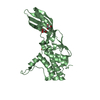
| Title | Crystal structure of SNX5-PX domain in P212121 space group | ||||||
 Components Components | Snx5 protein | ||||||
 Keywords Keywords | PROTEIN TRANSPORT / Sorting nexin / SNX5 / Phox / PhosphatidylInositol / PI(4 / 5)P2 | ||||||
| Function / homology |  Function and homology information Function and homology informationepidermal growth factor catabolic process / pinocytosis / cytoplasmic side of early endosome membrane / tubular endosome / macropinocytic cup / Golgi Associated Vesicle Biogenesis / phosphatidylinositol-5-phosphate binding / retromer complex / retrograde transport, endosome to Golgi / phosphatidylinositol-4-phosphate binding ...epidermal growth factor catabolic process / pinocytosis / cytoplasmic side of early endosome membrane / tubular endosome / macropinocytic cup / Golgi Associated Vesicle Biogenesis / phosphatidylinositol-5-phosphate binding / retromer complex / retrograde transport, endosome to Golgi / phosphatidylinositol-4-phosphate binding / phosphatidylinositol-3,5-bisphosphate binding / dynactin binding / phagocytic cup / brush border / D1 dopamine receptor binding / positive regulation of insulin receptor signaling pathway / ruffle / negative regulation of blood pressure / phosphatidylinositol binding / intracellular protein transport / cytoplasmic side of plasma membrane / endosome / perinuclear region of cytoplasm / positive regulation of DNA-templated transcription / cytosolSimilarity search - Function | ||||||
| Biological species |  Rattus norvegicus (Norway rat) Rattus norvegicus (Norway rat) | ||||||
| Method | X-RAY DIFFRACTION / SYNCHROTRON / MAD / Resolution: 2.19 Å | ||||||
 Authors Authors | Koharudin, L.M.I. / Furey, W. / Gronenborn, A.M. | ||||||
 Citation Citation | Journal: To be Published Title: The Phox domain of sorting nexin 5 lacks ptdins(3)p specificity and preferentially binds to ptdins(4,5)P2 Authors: Koharudin, L.M.I. / Furey, W. / Liu, H. / Liu, Y.-J. / Gronenborn, A.M. | ||||||
| History |
|
- Structure visualization
| Structure viewer | Molecule: MolmilJmol/JSmol |
|---|
- Downloads & links
-Download
| PDBx/mmCIF format | 3hpb.cif.gz | 43.6 KB | Display | PDBx/mmCIF format |
|---|---|---|---|---|
| PDB format | pdb3hpb.ent.gz | 33.8 KB | Display | PDB format |
| PDBx/mmJSON format | 3hpb.json.gz | Tree view | PDBx/mmJSON format | |
| Others |  Other downloads Other downloads |
-Validation report
| Arichive directory | https://data.pdbj.org/pub/pdb/validation_reports/hp/3hpbftp://data.pdbj.org/pub/pdb/validation_reports/hp/3hpb | HTTPS FTP |
|---|
-Related structure data


-Links
 PDBj
PDBj

- Assembly
| Deposited unit | 
| ||||||||
|---|---|---|---|---|---|---|---|---|---|
| 1 |
| ||||||||
| Unit cell |
|
-Components
| #1: Protein | Mass: 18649.512 Da / Num. of mol.: 1 / Fragment: Residues 20-180 Source method: isolated from a genetically manipulated source Source: (gene. exp.) Rattus norvegicus (Norway rat) / Gene: Snx5 / Plasmid: pET15b / Production host:  Escherichia coli (E. coli) / Strain (production host): BL21(DE3) / References: UniProt: B1H267 Escherichia coli (E. coli) / Strain (production host): BL21(DE3) / References: UniProt: B1H267 |
|---|---|
| #2: Chemical | ChemComp-CL / Chloride  Mass: 35.453 Da / Num. of mol.: 1 / Source method: obtained synthetically / Formula: Cl Mass: 35.453 Da / Num. of mol.: 1 / Source method: obtained synthetically / Formula: Cl |
| #3: Water | ChemComp-HOH / Water Mass: 18.015 Da / Num. of mol.: 135 / Source method: isolated from a natural source / Formula: H2O Mass: 18.015 Da / Num. of mol.: 135 / Source method: isolated from a natural source / Formula: H2O |
| Sequence details | THE ORIGINAL CONSTRUCT CONTAINED RESIDUES 1-180. THE FINAL CRYSTALLIZED PROTEIN CONSTRUCT SPANS ...THE ORIGINAL CONSTRUCT CONTAINED RESIDUES 1-180. THE FINAL CRYSTALLIZ |
-Experimental details
-Experiment
| Experiment | Method: X-RAY DIFFRACTION / Number of used crystals: 2 |
|---|
- Sample preparation
| Crystal | Density Matthews: 3.39 Å3/Da / Density % sol: 59.25 % |
|---|---|
| Crystal grow | Temperature: 277 K / pH: 8 Details: the protein solution in the final superdex-75 buffer (20 mM Tris.HCl, 100 mM NaCl, 0.02% NaN3, pH 8.0) was concentrated to 8 mg/ml. The Se-Met derivative and the 2.19 A crystals were ...Details: the protein solution in the final superdex-75 buffer (20 mM Tris.HCl, 100 mM NaCl, 0.02% NaN3, pH 8.0) was concentrated to 8 mg/ml. The Se-Met derivative and the 2.19 A crystals were obtained using an optimized initial crystallization condition of 8 ul of protein vs 1 ul of reservoir solution (0.2M (NH4)2SO4, 0.1M Sodium cacodylate tryhydrate pH 6.5, 30% PEG 8000) at 4C by sitting drop vapor diffusion, VAPOR DIFFUSION, SITTING DROP, temperature 277K |
-Data collection
| Diffraction |
| |||||||||||||||||||||
|---|---|---|---|---|---|---|---|---|---|---|---|---|---|---|---|---|---|---|---|---|---|---|
| Diffraction source |
| |||||||||||||||||||||
| Detector |
| |||||||||||||||||||||
| Radiation |
| |||||||||||||||||||||
| Radiation wavelength |
| |||||||||||||||||||||
| Reflection | Resolution: 2.19→28.78 Å / Num. obs: 12680 / % possible obs: 93.4 % / Observed criterion σ(I): 3 / Redundancy: 4.18 % / Rmerge(I) obs: 0.033 / Net I/σ(I): 28.1 | |||||||||||||||||||||
| Reflection shell | Resolution: 2.19→2.27 Å / Redundancy: 2.05 % / Rmerge(I) obs: 0.256 / Mean I/σ(I) obs: 3.5 / % possible all: 62.3 |

- Processing
| Software |
| ||||||||||||||||||||||||||||||||||||||||||||||||||||||||||||||||||||||||||||||||||||||||||||||||||||||||||||||||||||||||||||||||||||||||||||||||||||||||||||||||||||||||||
|---|---|---|---|---|---|---|---|---|---|---|---|---|---|---|---|---|---|---|---|---|---|---|---|---|---|---|---|---|---|---|---|---|---|---|---|---|---|---|---|---|---|---|---|---|---|---|---|---|---|---|---|---|---|---|---|---|---|---|---|---|---|---|---|---|---|---|---|---|---|---|---|---|---|---|---|---|---|---|---|---|---|---|---|---|---|---|---|---|---|---|---|---|---|---|---|---|---|---|---|---|---|---|---|---|---|---|---|---|---|---|---|---|---|---|---|---|---|---|---|---|---|---|---|---|---|---|---|---|---|---|---|---|---|---|---|---|---|---|---|---|---|---|---|---|---|---|---|---|---|---|---|---|---|---|---|---|---|---|---|---|---|---|---|---|---|---|---|---|---|---|---|
| Refinement | Method to determine structure: MAD / Resolution: 2.19→28.78 Å / Cor.coef. Fo:Fc: 0.945 / Cor.coef. Fo:Fc free: 0.917 / SU B: 6.449 / SU ML: 0.157 / Cross valid method: THROUGHOUT / σ(F): 0 / ESU R: 0.271 / ESU R Free: 0.232 / Stereochemistry target values: MAXIMUM LIKELIHOOD / Details: HYDROGENS HAVE BEEN ADDED IN THE RIDING POSITIONS
| ||||||||||||||||||||||||||||||||||||||||||||||||||||||||||||||||||||||||||||||||||||||||||||||||||||||||||||||||||||||||||||||||||||||||||||||||||||||||||||||||||||||||||
| Solvent computation | Ion probe radii: 0.8 Å / Shrinkage radii: 0.8 Å / VDW probe radii: 1.4 Å / Solvent model: MASK | ||||||||||||||||||||||||||||||||||||||||||||||||||||||||||||||||||||||||||||||||||||||||||||||||||||||||||||||||||||||||||||||||||||||||||||||||||||||||||||||||||||||||||
| Displacement parameters | Biso mean: 48.12 Å2
| ||||||||||||||||||||||||||||||||||||||||||||||||||||||||||||||||||||||||||||||||||||||||||||||||||||||||||||||||||||||||||||||||||||||||||||||||||||||||||||||||||||||||||
| Refinement step | Cycle: LAST / Resolution: 2.19→28.78 Å
| ||||||||||||||||||||||||||||||||||||||||||||||||||||||||||||||||||||||||||||||||||||||||||||||||||||||||||||||||||||||||||||||||||||||||||||||||||||||||||||||||||||||||||
| Refine LS restraints |
| ||||||||||||||||||||||||||||||||||||||||||||||||||||||||||||||||||||||||||||||||||||||||||||||||||||||||||||||||||||||||||||||||||||||||||||||||||||||||||||||||||||||||||
| LS refinement shell | Resolution: 2.19→2.25 Å / Total num. of bins used: 20
|
