 Movie
Movie Controller

[English] 日本語
 Yorodumi
- PDB-2fg9: CRYSTAL STRUCTURE OF A PUTATIVE 5-NITROIMIDAZOLE ANTIBIOTIC RESIS... -
+ Open data

- Basic information
| Entry | Database: PDB / ID: 2fg9 | ||||||
|---|---|---|---|---|---|---|---|
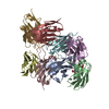
| Title | CRYSTAL STRUCTURE OF A PUTATIVE 5-NITROIMIDAZOLE ANTIBIOTIC RESISTANCE PROTEIN (BT_3078) FROM BACTEROIDES THETAIOTAOMICRON VPI-5482 AT 2.20 A RESOLUTION | ||||||
 Components Components | 5-nitroimidazole antibiotic resistance protein | ||||||
 Keywords Keywords |  OXIDOREDUCTASE / PUTATIVE 5-NITROIMIDAZOLE ANTIBIOTIC RESISTANCE PROTEIN / STRUCTURAL GENOMICS / JOINT CENTER FOR STRUCTURAL GENOMICS / JCSG / PROTEIN STRUCTURE INITIATIVE / PSI-2 OXIDOREDUCTASE / PUTATIVE 5-NITROIMIDAZOLE ANTIBIOTIC RESISTANCE PROTEIN / STRUCTURAL GENOMICS / JOINT CENTER FOR STRUCTURAL GENOMICS / JCSG / PROTEIN STRUCTURE INITIATIVE / PSI-2 | ||||||
| Function / homology |  Function and homology information Function and homology information | ||||||
| Biological species |  Bacteroides thetaiotaomicron (bacteria) Bacteroides thetaiotaomicron (bacteria) | ||||||
| Method | X-RAY DIFFRACTION / SYNCHROTRON / MAD / Resolution: 2.2 Å | ||||||
 Authors Authors | Joint Center for Structural Genomics (JCSG) | ||||||
 Citation Citation | Journal: To be published Title: Crystal structure of (np_811990.1) from BACTEROIDES THETAIOTAOMICRON VPI-5482 at 2.20 A resolution Authors: Joint Center for Structural Genomics (JCSG) | ||||||
| History |
| ||||||
| Remark 300 | BIOMOLECULE: 1 THIS ENTRY CONTAINS THE CRYSTALLOGRAPHIC ASYMMETRIC UNIT WHICH CONSISTS OF 1 CHAIN(S) ...BIOMOLECULE: 1 THIS ENTRY CONTAINS THE CRYSTALLOGRAPHIC ASYMMETRIC UNIT WHICH CONSISTS OF 1 CHAIN(S). SEE REMARK 350 FOR REMARK 300 INFORMATION ON GENERATING THE BIOLOGICAL MOLECULE(S). ASSIGNMENT OF THE BIOLOGICAL SUBUNIT AS A HEXAMER IS SUPPORTED BY DATA FROM SIZE EXCLUSION CHROMATOGRAPHY WITH STATIC LIGHT SCATTERING. HOWEVER, THE INTERACTIONS FORMING THE HEXAMER PRIMARILY INVOLVE THE EXPRESSION TAG, SO IT IS POSSIBLE THAT THE OLIGIMERIZATION STATE OF THE WILD-TYPE PROTEIN IS DIMERIC. |
- Structure visualization
| Structure viewer | Molecule: MolmilJmol/JSmol |
|---|
- Downloads & links
-Download
| PDBx/mmCIF format | 2fg9.cif.gz | 49.6 KB | Display | PDBx/mmCIF format |
|---|---|---|---|---|
| PDB format | pdb2fg9.ent.gz | 38 KB | Display | PDB format |
| PDBx/mmJSON format | 2fg9.json.gz | Tree view | PDBx/mmJSON format | |
| Others |  Other downloads Other downloads |
-Validation report
| Arichive directory | https://data.pdbj.org/pub/pdb/validation_reports/fg/2fg9ftp://data.pdbj.org/pub/pdb/validation_reports/fg/2fg9 | HTTPS FTP |
|---|
-Related structure data
| Similar structure data | |
|---|---|
| Other databases |


-Links
 PDBj
PDBj- Assembly
| Deposited unit | 
| ||||||||
|---|---|---|---|---|---|---|---|---|---|
| 1 | x 6
| ||||||||
| Unit cell |
| ||||||||
| Components on special symmetry positions |
| ||||||||
| Details | ASSIGNMENT OF THE BIOLOGICAL SUBUNIT AS A HEXAMER IS SUPPORTED BY DATA FROM SIZE EXCLUSION CHROMATOGRAPHY WITH STATIC LIGHT SCATTERING. HOWEVER, THE INTERACTIONS FORMING THE HEXAMER PRIMARILY INVOLVE THE EXPRESSION TAG, SO IT IS POSSIBLE THAT THE OLIGIMERIZATION STATE OF THE WILD-TYPE PROTEIN IS DIMERIC. |
-Components
| #1: Protein | Mass: 21013.602 Da / Num. of mol.: 1 Source method: isolated from a genetically manipulated source Source: (gene. exp.) Bacteroides thetaiotaomicron (bacteria)Strain: VPI-5482 / Gene: np_811990.1 / Production host: Escherichia coli (E. coli) / References: GenBank: 29340391, UniProt: Q8A377*PLUS |
|---|---|
| #2: Chemical | ChemComp-NI / Nickel  Mass: 58.693 Da / Num. of mol.: 1 / Source method: obtained synthetically / Formula: Ni Mass: 58.693 Da / Num. of mol.: 1 / Source method: obtained synthetically / Formula: Ni |
| #3: Chemical | ChemComp-FAD / Flavin adenine dinucleotide  Mass: 785.550 Da / Num. of mol.: 1 / Source method: obtained synthetically / Formula: C27H33N9O15P2 / Comment: FAD*YM Mass: 785.550 Da / Num. of mol.: 1 / Source method: obtained synthetically / Formula: C27H33N9O15P2 / Comment: FAD*YM |
| #4: Chemical | ChemComp-EDO / Ethylene glycol  Mass: 62.068 Da / Num. of mol.: 1 / Source method: obtained synthetically / Formula: C2H6O2 Mass: 62.068 Da / Num. of mol.: 1 / Source method: obtained synthetically / Formula: C2H6O2 |
| #5: Water | ChemComp-HOH / Water Mass: 18.015 Da / Num. of mol.: 46 / Source method: isolated from a natural source / Formula: H2O Mass: 18.015 Da / Num. of mol.: 46 / Source method: isolated from a natural source / Formula: H2O |
-Experimental details
-Experiment
| Experiment | Method: X-RAY DIFFRACTION / Number of used crystals: 1 |
|---|
- Sample preparation
| Crystal | Density Matthews: 2.49 Å3/Da / Density % sol: 50.21 % |
|---|---|
| Crystal grow | Temperature: 277 K / Method: vapor diffusion, sitting drop, nanodrop / pH: 7.9 Details: 0.2M (NH4)2HPO4, 20.0% PEG-3350, No Buffer, pH 7.9, VAPOR DIFFUSION,SITTING DROP,NANODROP, temperature 277K |
-Data collection
| Diffraction | Mean temperature: 100 K | |||||||||||||||||||||||||||||||||||||||||||||||||||||||||||||||||||||||||||||
|---|---|---|---|---|---|---|---|---|---|---|---|---|---|---|---|---|---|---|---|---|---|---|---|---|---|---|---|---|---|---|---|---|---|---|---|---|---|---|---|---|---|---|---|---|---|---|---|---|---|---|---|---|---|---|---|---|---|---|---|---|---|---|---|---|---|---|---|---|---|---|---|---|---|---|---|---|---|---|
| Diffraction source | Source: SYNCHROTRON / Site: ALS  / Beamline: 8.2.1 / Wavelength: 0.9798, 0.9797, 1.0163 / Beamline: 8.2.1 / Wavelength: 0.9798, 0.9797, 1.0163 | |||||||||||||||||||||||||||||||||||||||||||||||||||||||||||||||||||||||||||||
| Detector | Type: ADSC QUANTUM 210 / Detector: CCD / Date: Sep 28, 2005 | |||||||||||||||||||||||||||||||||||||||||||||||||||||||||||||||||||||||||||||
| Radiation | Monochromator: Double Crystal Si(111) / Protocol: MAD / Monochromatic (M) / Laue (L): M / Scattering type: x-ray | |||||||||||||||||||||||||||||||||||||||||||||||||||||||||||||||||||||||||||||
| Radiation wavelength |
| |||||||||||||||||||||||||||||||||||||||||||||||||||||||||||||||||||||||||||||
| Reflection | Resolution: 2.2→28.57 Å / Num. obs: 10216 / % possible obs: 98.2 % / Redundancy: 11.2 % / Rmerge(I) obs: 0.068 / Net I/σ(I): 22.58 | |||||||||||||||||||||||||||||||||||||||||||||||||||||||||||||||||||||||||||||
| Reflection shell | Rmerge(I) obs: 0.755 / Diffraction-ID: 1
|
-Phasing
| Phasing | Method: MAD |
|---|
- Processing
| Software |
| |||||||||||||||||||||||||||||||||||||||||||||||||||||||||||||||||||||||||||||||||||||||||||||||||||||||||||||||||||||||||||||
|---|---|---|---|---|---|---|---|---|---|---|---|---|---|---|---|---|---|---|---|---|---|---|---|---|---|---|---|---|---|---|---|---|---|---|---|---|---|---|---|---|---|---|---|---|---|---|---|---|---|---|---|---|---|---|---|---|---|---|---|---|---|---|---|---|---|---|---|---|---|---|---|---|---|---|---|---|---|---|---|---|---|---|---|---|---|---|---|---|---|---|---|---|---|---|---|---|---|---|---|---|---|---|---|---|---|---|---|---|---|---|---|---|---|---|---|---|---|---|---|---|---|---|---|---|---|---|
| Refinement | Method to determine structure: MAD / Resolution: 2.2→28.57 Å / Cor.coef. Fo:Fc: 0.94 / Cor.coef. Fo:Fc free: 0.943 / SU B: 14.012 / SU ML: 0.179 / TLS residual ADP flag: LIKELY RESIDUAL / Cross valid method: THROUGHOUT / ESU R: 0.283 / ESU R Free: 0.206 Stereochemistry target values: MAXIMUM LIKELIHOOD WITH PHASES Details: 1. HYDROGENS HAVE BEEN ADDED IN THE RIDING POSITIONS 2. DENSITY FOR RESIDUES 81-86 AND 158-159 IS DISORDERED AND NOT MODELED. 3. ASSIGNMENT OF NI IS BASED ON COORDINATION NUMBER AND GEOMETRY. ...Details: 1. HYDROGENS HAVE BEEN ADDED IN THE RIDING POSITIONS 2. DENSITY FOR RESIDUES 81-86 AND 158-159 IS DISORDERED AND NOT MODELED. 3. ASSIGNMENT OF NI IS BASED ON COORDINATION NUMBER AND GEOMETRY. 4. A MET-INHIBITION PROTOCOL WAS USED FOR SELENOMETHIONINE INCORPORATION DURING PROTEIN EXPRESSION. THE OCCUPANCY OF THE SE ATOMS IN THE MSE RESIDUES WAS REDUCED TO 0.75 TO ACCOUNT FOR THE REDUCED SCATTERING POWER DUE TO PARTIAL S-MET INCORPORATION.
| |||||||||||||||||||||||||||||||||||||||||||||||||||||||||||||||||||||||||||||||||||||||||||||||||||||||||||||||||||||||||||||
| Solvent computation | Ion probe radii: 0.8 Å / Shrinkage radii: 0.8 Å / VDW probe radii: 1.2 Å / Solvent model: MASK | |||||||||||||||||||||||||||||||||||||||||||||||||||||||||||||||||||||||||||||||||||||||||||||||||||||||||||||||||||||||||||||
| Displacement parameters | Biso mean: 47.821 Å2 | |||||||||||||||||||||||||||||||||||||||||||||||||||||||||||||||||||||||||||||||||||||||||||||||||||||||||||||||||||||||||||||
| Refinement step | Cycle: LAST / Resolution: 2.2→28.57 Å
| |||||||||||||||||||||||||||||||||||||||||||||||||||||||||||||||||||||||||||||||||||||||||||||||||||||||||||||||||||||||||||||
| Refine LS restraints |
| |||||||||||||||||||||||||||||||||||||||||||||||||||||||||||||||||||||||||||||||||||||||||||||||||||||||||||||||||||||||||||||
| LS refinement shell | Resolution: 2.2→2.258 Å / Total num. of bins used: 20
| |||||||||||||||||||||||||||||||||||||||||||||||||||||||||||||||||||||||||||||||||||||||||||||||||||||||||||||||||||||||||||||
| Refinement TLS params. | Method: refined / Origin x: 40.3046 Å / Origin y: 15.5919 Å / Origin z: 49.4336 Å
|
