 Movie
Movie Controller

[English] 日本語
 Yorodumi
- PDB-3hlz: Crystal structure of BT_1490 (NP_810393.1) from BACTEROIDES THETA... -
+ Open data

- Basic information
| Entry | Database: PDB / ID: 3hlz | ||||||
|---|---|---|---|---|---|---|---|
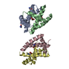
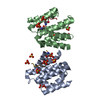
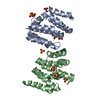
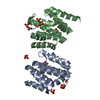
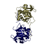
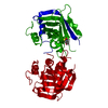
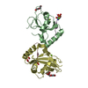
| Title | Crystal structure of BT_1490 (NP_810393.1) from BACTEROIDES THETAIOTAOMICRON VPI-5482 at 1.50 A resolution | ||||||
 Components Components | uncharacterized protein BT_1490 | ||||||
 Keywords Keywords |  structural genomics / unknown function / NP_810393.1 / BT_1490 / Joint Center for Structural Genomics / JCSG / Protein Structure Initiative / PSI-2 structural genomics / unknown function / NP_810393.1 / BT_1490 / Joint Center for Structural Genomics / JCSG / Protein Structure Initiative / PSI-2 | ||||||
| Function / homology |  Function and homology information Function and homology informationFour Helix Bundle (Hemerythrin (Met), subunit A) - #1090 / Domain of unknown function DUF3806 / Domain of unknown function DUF3805 / Domain of unknown function (DUF3805) / Domain of unknown function (DUF3806) / Mog1/PsbP, alpha/beta/alpha sandwich / Protein Transport Mog1p; Chain A / Mog1/PsbP, alpha/beta/alpha sandwich / Four Helix Bundle (Hemerythrin (Met), subunit A) / Up-down Bundle ...Four Helix Bundle (Hemerythrin (Met), subunit A) - #1090 / Domain of unknown function DUF3806 / Domain of unknown function DUF3805 / Domain of unknown function (DUF3805) / Domain of unknown function (DUF3806) / Mog1/PsbP, alpha/beta/alpha sandwich / Protein Transport Mog1p; Chain A / Mog1/PsbP, alpha/beta/alpha sandwich / Four Helix Bundle (Hemerythrin (Met), subunit A) / Up-down Bundle / 3-Layer(aba) Sandwich / Mainly Alpha / Alpha Beta Similarity search - Domain/homology | ||||||
| Biological species |  Bacteroides thetaiotaomicron VPI-5482 (bacteria) Bacteroides thetaiotaomicron VPI-5482 (bacteria) | ||||||
| Method | X-RAY DIFFRACTION / SYNCHROTRON / MAD / Resolution: 1.5 Å | ||||||
 Authors Authors | Joint Center for Structural Genomics (JCSG) | ||||||
 Citation Citation | Journal: To be Published Title: Crystal structure of BT_1490 (NP_810393.1) from BACTEROIDES THETAIOTAOMICRON VPI-5482 at 1.50 A resolution Authors: Joint Center for Structural Genomics (JCSG) | ||||||
| History |
|
- Structure visualization
| Structure viewer | Molecule: MolmilJmol/JSmol |
|---|
- Downloads & links
-Download
| PDBx/mmCIF format | 3hlz.cif.gz | 129.4 KB | Display | PDBx/mmCIF format |
|---|---|---|---|---|
| PDB format | pdb3hlz.ent.gz | 103.8 KB | Display | PDB format |
| PDBx/mmJSON format | 3hlz.json.gz | Tree view | PDBx/mmJSON format | |
| Others |  Other downloads Other downloads |
-Validation report
| Arichive directory | https://data.pdbj.org/pub/pdb/validation_reports/hl/3hlzftp://data.pdbj.org/pub/pdb/validation_reports/hl/3hlz | HTTPS FTP |
|---|
-Related structure data
| Similar structure data | |
|---|---|
| Other databases |

-Links
 PDBj
PDBj
- Assembly
| Deposited unit | 
| ||||||||
|---|---|---|---|---|---|---|---|---|---|
| 1 | 
| ||||||||
| 2 | 
| ||||||||
| Unit cell |
|
-Components
| #1: Protein | Mass: 30623.516 Da / Num. of mol.: 2 Source method: isolated from a genetically manipulated source Source: (gene. exp.) Bacteroides thetaiotaomicron VPI-5482 (bacteria)Gene: BT_1480, NP_810393.1 / Plasmid: SpeedET / Production host: Escherichia Coli (E. coli) / Strain (production host): HK100 / References: UniProt: Q8A7P4#2: Chemical | ChemComp-GOL / Glycerol  Mass: 92.094 Da / Num. of mol.: 4 / Source method: obtained synthetically / Formula: C3H8O3 Mass: 92.094 Da / Num. of mol.: 4 / Source method: obtained synthetically / Formula: C3H8O3#3: Chemical | ChemComp-ACT / Acetate  Mass: 59.044 Da / Num. of mol.: 4 / Source method: obtained synthetically / Formula: C2H3O2 Mass: 59.044 Da / Num. of mol.: 4 / Source method: obtained synthetically / Formula: C2H3O2#4: Chemical | ChemComp-CA / |   Mass: 40.078 Da / Num. of mol.: 1 / Source method: obtained synthetically / Formula: Ca Mass: 40.078 Da / Num. of mol.: 1 / Source method: obtained synthetically / Formula: Ca#5: Water | ChemComp-HOH / | Water Mass: 18.015 Da / Num. of mol.: 448 / Source method: isolated from a natural source / Formula: H2O Mass: 18.015 Da / Num. of mol.: 448 / Source method: isolated from a natural source / Formula: H2OSequence details | SEQUENCE THE CONSTRUCT (RESIDUES 23-178) WAS EXPRESSED WITH A PURIFICATION TAG MGSDKIHHHHHHENLYFQG. ...SEQUENCE THE CONSTRUCT (RESIDUES 23-178) WAS EXPRESSED WITH A PURIFICATI | |
|---|
-Experimental details
-Experiment
| Experiment | Method: X-RAY DIFFRACTION / Number of used crystals: 1 |
|---|
- Sample preparation
| Crystal | Density Matthews: 2.15 Å3/Da / Density % sol: 42.8 % |
|---|---|
| Crystal grow | Temperature: 277 K / Method: vapor diffusion, sitting drop / pH: 6 Details: 0.2000M Ca(OAc)2, 20.0000% PEG-8000, 0.1M MES pH 6.0, NANODROP, VAPOR DIFFUSION, SITTING DROP, temperature 277K |
-Data collection
| Diffraction | Mean temperature: 100 K | |||||||||||||||||||||||||||||||||||||||||||||||||||||||||||||||||||||||||||||
|---|---|---|---|---|---|---|---|---|---|---|---|---|---|---|---|---|---|---|---|---|---|---|---|---|---|---|---|---|---|---|---|---|---|---|---|---|---|---|---|---|---|---|---|---|---|---|---|---|---|---|---|---|---|---|---|---|---|---|---|---|---|---|---|---|---|---|---|---|---|---|---|---|---|---|---|---|---|---|
| Diffraction source | Source: SYNCHROTRON / Site: SSRL  / Beamline: BL11-1 / Wavelength: 0.91837,0.97845 / Beamline: BL11-1 / Wavelength: 0.91837,0.97845 | |||||||||||||||||||||||||||||||||||||||||||||||||||||||||||||||||||||||||||||
| Detector | Type: MARMOSAIC 325 mm CCD / Detector: CCD / Date: Apr 16, 2009 / Details: Flat mirror (vertical focusing) | |||||||||||||||||||||||||||||||||||||||||||||||||||||||||||||||||||||||||||||
| Radiation | Monochromator: Single crystal Si(111) bent monochromator (horizontal focusing) Protocol: MAD / Monochromatic (M) / Laue (L): M / Scattering type: x-ray | |||||||||||||||||||||||||||||||||||||||||||||||||||||||||||||||||||||||||||||
| Radiation wavelength |
| |||||||||||||||||||||||||||||||||||||||||||||||||||||||||||||||||||||||||||||
| Reflection | Resolution: 1.5→29.814 Å / Num. obs: 76979 / % possible obs: 87.9 % / Observed criterion σ(I): -3 / Biso Wilson estimate: 15.871 Å2 / Rmerge(I) obs: 0.039 / Net I/σ(I): 10.1 | |||||||||||||||||||||||||||||||||||||||||||||||||||||||||||||||||||||||||||||
| Reflection shell |
|
-Phasing
| Phasing | Method: MAD |
|---|
- Processing
| Software |
| |||||||||||||||||||||||||||||||||||||||||||||||||||||||||||||||||||||||||||||||||||||||||||||||||||||||||||||||||||||||||||||||||||||||
|---|---|---|---|---|---|---|---|---|---|---|---|---|---|---|---|---|---|---|---|---|---|---|---|---|---|---|---|---|---|---|---|---|---|---|---|---|---|---|---|---|---|---|---|---|---|---|---|---|---|---|---|---|---|---|---|---|---|---|---|---|---|---|---|---|---|---|---|---|---|---|---|---|---|---|---|---|---|---|---|---|---|---|---|---|---|---|---|---|---|---|---|---|---|---|---|---|---|---|---|---|---|---|---|---|---|---|---|---|---|---|---|---|---|---|---|---|---|---|---|---|---|---|---|---|---|---|---|---|---|---|---|---|---|---|---|---|
| Refinement | Method to determine structure: MAD / Resolution: 1.5→29.814 Å / Cor.coef. Fo:Fc: 0.965 / Cor.coef. Fo:Fc free: 0.957 / Occupancy max: 1 / Occupancy min: 0.3 / SU B: 2.953 / SU ML: 0.055 / TLS residual ADP flag: LIKELY RESIDUAL / Cross valid method: THROUGHOUT / σ(F): 0 / ESU R: 0.079 / ESU R Free: 0.078 Stereochemistry target values: MAXIMUM LIKELIHOOD WITH PHASES Details: 1. HYDROGENS HAVE BEEN ADDED IN THE RIDING POSITIONS. 2. A MET-INHIBITION PROTOCOL WAS USED FOR SELENOMETHIONINE INCORPORATION DURING PROTEIN EXPRESSION. THE OCCUPANCY OF THE SE ATOMS IN THE ...Details: 1. HYDROGENS HAVE BEEN ADDED IN THE RIDING POSITIONS. 2. A MET-INHIBITION PROTOCOL WAS USED FOR SELENOMETHIONINE INCORPORATION DURING PROTEIN EXPRESSION. THE OCCUPANCY OF THE SE ATOMS IN THE MSE RESIDUES WAS REDUCED TO 0.85 FOR THE REDUCED SCATTERING POWER DUE TO PARTIAL S-MET INCORPORATION. 3. ATOM RECORDS CONTAIN RESIDUAL B FACTORS ONLY. 4 GLYCEROL (GOL), ACETATE (ACT), AND CALCIUM (CA) FROM THE CRYSTALLIZATION/CRYOGEN SOLUTIONS WERE MODELED INTO THE STRUCTURE. 4. UN-ASSIGNED ELECTRON DENSITES NEAR THE SIDECHAINS OF GLU 177 AND GLU 213 ON THE A SUBUNIT, AND LYS A73 ON THE A SUBUNIT WERE NOT MODELED.
| |||||||||||||||||||||||||||||||||||||||||||||||||||||||||||||||||||||||||||||||||||||||||||||||||||||||||||||||||||||||||||||||||||||||
| Solvent computation | Ion probe radii: 0.8 Å / Shrinkage radii: 0.8 Å / VDW probe radii: 1.2 Å / Solvent model: BABINET MODEL WITH MASK | |||||||||||||||||||||||||||||||||||||||||||||||||||||||||||||||||||||||||||||||||||||||||||||||||||||||||||||||||||||||||||||||||||||||
| Displacement parameters | Biso max: 97.76 Å2 / Biso mean: 24.816 Å2 / Biso min: 11.59 Å2
| |||||||||||||||||||||||||||||||||||||||||||||||||||||||||||||||||||||||||||||||||||||||||||||||||||||||||||||||||||||||||||||||||||||||
| Refinement step | Cycle: LAST / Resolution: 1.5→29.814 Å
| |||||||||||||||||||||||||||||||||||||||||||||||||||||||||||||||||||||||||||||||||||||||||||||||||||||||||||||||||||||||||||||||||||||||
| Refine LS restraints |
| |||||||||||||||||||||||||||||||||||||||||||||||||||||||||||||||||||||||||||||||||||||||||||||||||||||||||||||||||||||||||||||||||||||||
| LS refinement shell | Resolution: 1.5→1.539 Å / Total num. of bins used: 20
| |||||||||||||||||||||||||||||||||||||||||||||||||||||||||||||||||||||||||||||||||||||||||||||||||||||||||||||||||||||||||||||||||||||||
| Refinement TLS params. | Method: refined / Refine-ID: X-RAY DIFFRACTION
| |||||||||||||||||||||||||||||||||||||||||||||||||||||||||||||||||||||||||||||||||||||||||||||||||||||||||||||||||||||||||||||||||||||||
| Refinement TLS group |
|
