 Movie
Movie Controller

[English] 日本語
 Yorodumi
- PDB-3hkl: Crystal Structure of the Frizzled-like Cysteine-rich Domain of MuSK -
+ Open data

- Basic information
| Entry | Database: PDB / ID: 3hkl | |||||||||
|---|---|---|---|---|---|---|---|---|---|---|
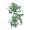
| Title | Crystal Structure of the Frizzled-like Cysteine-rich Domain of MuSK | |||||||||
 Components Components | Muscle, skeletal receptor tyrosine protein kinase Skeletal muscle Skeletal muscle | |||||||||
 Keywords Keywords | Transferase / Signaling protein / MuSK / RECEPTOR TYROSINE KINASE / FRIZZLED CRD / ATP-binding / Disulfide bond / Glycoprotein / Immunoglobulin domain / Kinase / Membrane / Nucleotide-binding / Phosphoprotein / Receptor / Transmembrane / Tyrosine-protein kinase | |||||||||
| Function / homology |  Function and homology information Function and homology informationpositive regulation of protein geranylgeranylation / regulation of synaptic assembly at neuromuscular junction / positive regulation of synaptic assembly at neuromuscular junction / positive regulation of motor neuron apoptotic process / skeletal muscle acetylcholine-gated channel clustering / positive regulation of synaptic transmission, cholinergic / positive regulation of skeletal muscle acetylcholine-gated channel clustering / Wnt-protein binding / response to muscle activity / motor neuron apoptotic process ...positive regulation of protein geranylgeranylation / regulation of synaptic assembly at neuromuscular junction / positive regulation of synaptic assembly at neuromuscular junction / positive regulation of motor neuron apoptotic process / skeletal muscle acetylcholine-gated channel clustering / positive regulation of synaptic transmission, cholinergic / positive regulation of skeletal muscle acetylcholine-gated channel clustering / Wnt-protein binding / response to muscle activity / motor neuron apoptotic process / neuromuscular junction development / cochlea development / enzyme-linked receptor protein signaling pathway / receptor clustering / positive regulation of Rac protein signal transduction / response to axon injury / response to electrical stimulus / transmembrane receptor protein tyrosine kinase activity / cell projection / PDZ domain binding / long-term synaptic potentiation / cell surface receptor protein tyrosine kinase signaling pathway / neuromuscular junction / receptor protein-tyrosine kinase / memory / positive regulation of neuron projection development / positive regulation of peptidyl-tyrosine phosphorylation / retina development in camera-type eye / postsynaptic membrane / protein tyrosine kinase activity / positive regulation of phosphatidylinositol 3-kinase/protein kinase B signal transduction / cell differentiation / receptor complex / positive regulation of protein phosphorylation / phosphorylation / external side of plasma membrane / negative regulation of gene expression / synapse / regulation of DNA-templated transcription / positive regulation of gene expression / protein kinase binding / ATP binding / membrane / metal ion binding / plasma membraneSimilarity search - Function | |||||||||
| Biological species |  Rattus norvegicus (Norway rat) Rattus norvegicus (Norway rat) | |||||||||
| Method | X-RAY DIFFRACTION / SYNCHROTRON / SAD / Resolution: 2.1 Å | |||||||||
 Authors Authors | Stiegler, A.L. / Hubbard, S.R. | |||||||||
 Citation Citation | Journal: J.Mol.Biol. / Year: 2009 Title: Crystal structure of the frizzled-like cysteine-rich domain of the receptor tyrosine kinase MuSK. Authors: Stiegler, A.L. / Burden, S.J. / Hubbard, S.R. | |||||||||
| History |
|
- Structure visualization
| Structure viewer | Molecule: MolmilJmol/JSmol |
|---|
- Downloads & links
-Download
| PDBx/mmCIF format | 3hkl.cif.gz | 68.3 KB | Display | PDBx/mmCIF format |
|---|---|---|---|---|
| PDB format | pdb3hkl.ent.gz | 53 KB | Display | PDB format |
| PDBx/mmJSON format | 3hkl.json.gz | Tree view | PDBx/mmJSON format | |
| Others |  Other downloads Other downloads |
-Validation report
| Arichive directory | https://data.pdbj.org/pub/pdb/validation_reports/hk/3hklftp://data.pdbj.org/pub/pdb/validation_reports/hk/3hkl | HTTPS FTP |
|---|
-Related structure data
| Similar structure data |
|---|


-Links
 PDBj
PDBj

- Assembly
| Deposited unit | 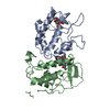
| ||||||||
|---|---|---|---|---|---|---|---|---|---|
| 1 |
| ||||||||
| Unit cell |
|
-Components
| #1: Protein | Skeletal muscle / Muscle-specific kinase receptor / MuSK Mass: 21888.062 Da / Num. of mol.: 2 / Fragment: FZ-CRD, residues 313-494 Source method: isolated from a genetically manipulated source Source: (gene. exp.) Rattus norvegicus (Norway rat) / Gene: Musk / Plasmid: PACGP67 / Production host:   Spodoptera frugiperda (fall armyworm) Spodoptera frugiperda (fall armyworm)References: UniProt: Q62838, receptor protein-tyrosine kinase#2: Polysaccharide | / Mass: 424.401 Da / Num. of mol.: 2Source method: isolated from a genetically manipulated source #3: Water | ChemComp-HOH / | Water Mass: 18.015 Da / Num. of mol.: 63 / Source method: isolated from a natural source / Formula: H2O Mass: 18.015 Da / Num. of mol.: 63 / Source method: isolated from a natural source / Formula: H2O |
|---|
-Experimental details
-Experiment
| Experiment | Method: X-RAY DIFFRACTION / Number of used crystals: 1 |
|---|
- Sample preparation
| Crystal | Density Matthews: 2.2 Å3/Da / Density % sol: 44.02 % |
|---|---|
| Crystal grow | Temperature: 293 K / Method: vapor diffusion, hanging drop / pH: 8.5 Details: 5% PEG 4000, 0.2M SODIUM ACETATE, 0.1M TRIS PH 8.5, 5% GLYCEROL, VAPOR DIFFUSION, HANGING DROP, temperature 293K |
-Data collection
| Diffraction | Mean temperature: 100 K |
|---|---|
| Diffraction source | Source: SYNCHROTRON / Site: NSLS  / Beamline: X4C / Wavelength: 0.9787 Å / Beamline: X4C / Wavelength: 0.9787 Å |
| Detector | Type: MAR scanner 345 mm plate / Detector: IMAGE PLATE / Date: Sep 29, 2006 |
| Radiation | Protocol: SINGLE WAVELENGTH / Monochromatic (M) / Laue (L): M / Scattering type: x-ray |
| Radiation wavelength | Wavelength: 0.9787 Å / Relative weight: 1 |
| Reflection | Resolution: 2.1→34.47 Å / Num. all: 22374 / Num. obs: 22050 |
| Reflection shell | Resolution: 2.1→2.18 Å / Redundancy: 2.8 % / Mean I/σ(I) obs: 4.5 / Rsym value: 0.299 / % possible all: 96.9 |
- Processing
| Software |
| ||||||||||||||||||||||||||||||||||||||||||||||||||||||||||||||||||||||||||||||||||||||||||||||||||||||||||||||||||||||||||||||||||||||||||||||||||||||||||||||||||||||||||
|---|---|---|---|---|---|---|---|---|---|---|---|---|---|---|---|---|---|---|---|---|---|---|---|---|---|---|---|---|---|---|---|---|---|---|---|---|---|---|---|---|---|---|---|---|---|---|---|---|---|---|---|---|---|---|---|---|---|---|---|---|---|---|---|---|---|---|---|---|---|---|---|---|---|---|---|---|---|---|---|---|---|---|---|---|---|---|---|---|---|---|---|---|---|---|---|---|---|---|---|---|---|---|---|---|---|---|---|---|---|---|---|---|---|---|---|---|---|---|---|---|---|---|---|---|---|---|---|---|---|---|---|---|---|---|---|---|---|---|---|---|---|---|---|---|---|---|---|---|---|---|---|---|---|---|---|---|---|---|---|---|---|---|---|---|---|---|---|---|---|---|---|
| Refinement | Method to determine structure: SAD / Resolution: 2.1→34.47 Å / Cor.coef. Fo:Fc: 0.939 / Cor.coef. Fo:Fc free: 0.922 / SU B: 4.892 / SU ML: 0.134 / Cross valid method: THROUGHOUT / ESU R: 0.22 / ESU R Free: 0.191 / Stereochemistry target values: MAXIMUM LIKELIHOOD / Details: HYDROGENS HAVE BEEN ADDED IN THE RIDING POSITIONS
| ||||||||||||||||||||||||||||||||||||||||||||||||||||||||||||||||||||||||||||||||||||||||||||||||||||||||||||||||||||||||||||||||||||||||||||||||||||||||||||||||||||||||||
| Solvent computation | Ion probe radii: 0.8 Å / Shrinkage radii: 0.8 Å / VDW probe radii: 1.2 Å / Solvent model: MASK | ||||||||||||||||||||||||||||||||||||||||||||||||||||||||||||||||||||||||||||||||||||||||||||||||||||||||||||||||||||||||||||||||||||||||||||||||||||||||||||||||||||||||||
| Displacement parameters | Biso mean: 37.338 Å2
| ||||||||||||||||||||||||||||||||||||||||||||||||||||||||||||||||||||||||||||||||||||||||||||||||||||||||||||||||||||||||||||||||||||||||||||||||||||||||||||||||||||||||||
| Refinement step | Cycle: LAST / Resolution: 2.1→34.47 Å
| ||||||||||||||||||||||||||||||||||||||||||||||||||||||||||||||||||||||||||||||||||||||||||||||||||||||||||||||||||||||||||||||||||||||||||||||||||||||||||||||||||||||||||
| Refine LS restraints |
| ||||||||||||||||||||||||||||||||||||||||||||||||||||||||||||||||||||||||||||||||||||||||||||||||||||||||||||||||||||||||||||||||||||||||||||||||||||||||||||||||||||||||||
| LS refinement shell | Resolution: 2.1→2.155 Å / Total num. of bins used: 20
|
