 Movie
Movie Controller

[English] 日本語
 Yorodumi
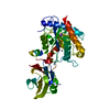
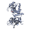
- PDB-3d5n: Crystal structure of the Q97W15_SULSO protein from Sulfolobus sol... -
+ Open data

- Basic information
| Entry | Database: PDB / ID: 3d5n | ||||||
|---|---|---|---|---|---|---|---|
| Title | Crystal structure of the Q97W15_SULSO protein from Sulfolobus solfataricus. NESG target SsR125. | ||||||
 Components Components | Q97W15_SULSO | ||||||
 Keywords Keywords |  structural genomics / unknown function / Q97W15 / SULSO / NESG / SsR125 / PSI-2 / Protein Structure Initiative / Northeast Structural Genomics Consortium structural genomics / unknown function / Q97W15 / SULSO / NESG / SsR125 / PSI-2 / Protein Structure Initiative / Northeast Structural Genomics Consortium | ||||||
| Function / homology | MobA-like NTP transferase / MobA-like NTP transferase domain / Spore Coat Polysaccharide Biosynthesis Protein SpsA; Chain A / Spore Coat Polysaccharide Biosynthesis Protein SpsA; Chain A / Nucleotide-diphospho-sugar transferases / Alpha-Beta Complex / Alpha Beta / NTP_transf_3 domain-containing protein Function and homology information Function and homology information | ||||||
| Biological species |   Sulfolobus solfataricus (archaea) Sulfolobus solfataricus (archaea) | ||||||
| Method | X-RAY DIFFRACTION / SYNCHROTRON / MAD / Resolution: 2.8 Å | ||||||
 Authors Authors | Vorobiev, S.M. / Chen, Y. / Seetharaman, J. / Lee, D. / Foote, R.E. / Maglaqui, M. / Janjua, H. / Xiao, R. / Acton, T.B. / Montelione, G.T. ...Vorobiev, S.M. / Chen, Y. / Seetharaman, J. / Lee, D. / Foote, R.E. / Maglaqui, M. / Janjua, H. / Xiao, R. / Acton, T.B. / Montelione, G.T. / Tong, L. / Hunt, J.F. / Northeast Structural Genomics Consortium (NESG) | ||||||
 Citation Citation | Journal: To be Published Title: Crystal structure of the Q97W15_SULSO protein from Sulfolobus solfataricus. NESG target SsR125. Authors: Vorobiev, S.M. / Chen, Y. / Seetharaman, J. / Lee, D. / Foote, R.E. / Maglaqui, M. / Janjua, H. / Xiao, R. / Acton, T.B. / Montelione, G.T. / Tong, L. / Hunt, J.F. | ||||||
| History |
|
- Structure visualization
| Structure viewer | Molecule: MolmilJmol/JSmol |
|---|
- Downloads & links
-Download
| PDBx/mmCIF format | 3d5n.cif.gz | 263 KB | Display | PDBx/mmCIF format |
|---|---|---|---|---|
| PDB format | pdb3d5n.ent.gz | 222.2 KB | Display | PDB format |
| PDBx/mmJSON format | 3d5n.json.gz | Tree view | PDBx/mmJSON format | |
| Others |  Other downloads Other downloads |
-Validation report
| Arichive directory | https://data.pdbj.org/pub/pdb/validation_reports/d5/3d5nftp://data.pdbj.org/pub/pdb/validation_reports/d5/3d5n | HTTPS FTP |
|---|
-Related structure data
| Similar structure data | |
|---|---|
| Other databases |


-Links
 PDBj
PDBj

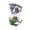
- Assembly
| Deposited unit | 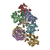
| ||||||||
|---|---|---|---|---|---|---|---|---|---|
| 1 | 
| ||||||||
| 2 | 
| ||||||||
| 3 | 
| ||||||||
| 4 | 
| ||||||||
| Unit cell |
| ||||||||
| Details | authors state that the biological unit is dimer according to gel-filtration experiments. |
-Components
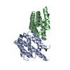
| #1: Protein | Mass: 22757.844 Da / Num. of mol.: 8 Source method: isolated from a genetically manipulated source Source: (gene. exp.) Sulfolobus solfataricus (archaea) / Strain: ATCC 35092 / DSM 1617 / JCM 11322 / P2 / Gene: SSO2432 / Plasmid: pET 21-23C / Production host:  Escherichia coli (E. coli) / Strain (production host): BL21(DE3) + Magic / References: UniProt: Q97W15 Escherichia coli (E. coli) / Strain (production host): BL21(DE3) + Magic / References: UniProt: Q97W15#2: Water | ChemComp-HOH / | Water Mass: 18.015 Da / Num. of mol.: 33 / Source method: isolated from a natural source / Formula: H2O Mass: 18.015 Da / Num. of mol.: 33 / Source method: isolated from a natural source / Formula: H2O |
|---|
-Experimental details
-Experiment
| Experiment | Method: X-RAY DIFFRACTION / Number of used crystals: 1 |
|---|
- Sample preparation
| Crystal | Density Matthews: 3.58 Å3/Da / Density % sol: 65.68 % |
|---|---|
| Crystal grow | Temperature: 291 K / Method: vapor diffusion, hanging drop Details: 1.8 - 2.0 M sodium malonate, VAPOR DIFFUSION, HANGING DROP, temperature 291K |
-Data collection
| Diffraction | Mean temperature: 100 K | ||||||||||||
|---|---|---|---|---|---|---|---|---|---|---|---|---|---|
| Diffraction source | Source: SYNCHROTRON / Site: NSLS  / Beamline: X4A / Wavelength: 0.97904, 0.97922, 0.96786 / Beamline: X4A / Wavelength: 0.97904, 0.97922, 0.96786 | ||||||||||||
| Detector | Type: ADSC QUANTUM 4 / Detector: CCD / Date: Apr 17, 2008 | ||||||||||||
| Radiation | Protocol: MAD / Monochromatic (M) / Laue (L): M / Scattering type: x-ray | ||||||||||||
| Radiation wavelength |
| ||||||||||||
| Reflection | Resolution: 2.8→50 Å / Num. all: 127020 / Num. obs: 126385 / % possible obs: 99.5 % / Observed criterion σ(F): 0 / Observed criterion σ(I): 0 / Redundancy: 7.4 % / Biso Wilson estimate: 35.3 Å2 / Rmerge(I) obs: 0.088 / Net I/σ(I): 26.3 | ||||||||||||
| Reflection shell | Resolution: 2.8→2.9 Å / Redundancy: 6.8 % / Rmerge(I) obs: 0.986 / Mean I/σ(I) obs: 1.85 / Num. unique all: 12662 / % possible all: 100 |
- Processing
| Software |
| ||||||||||||||||||||||||||||||||||||||||||||||||||||||||||||
|---|---|---|---|---|---|---|---|---|---|---|---|---|---|---|---|---|---|---|---|---|---|---|---|---|---|---|---|---|---|---|---|---|---|---|---|---|---|---|---|---|---|---|---|---|---|---|---|---|---|---|---|---|---|---|---|---|---|---|---|---|---|
| Refinement | Method to determine structure: MAD / Resolution: 2.8→48.47 Å / Rfactor Rfree error: 0.004 / Data cutoff high absF: 278545.18 / Data cutoff low absF: 0 / Isotropic thermal model: RESTRAINED / Cross valid method: THROUGHOUT / σ(F): 2 / Stereochemistry target values: Engh & Huber
| ||||||||||||||||||||||||||||||||||||||||||||||||||||||||||||
| Solvent computation | Solvent model: FLAT MODEL / Bsol: 58.1558 Å2 / ksol: 0.360337 e/Å3 | ||||||||||||||||||||||||||||||||||||||||||||||||||||||||||||
| Displacement parameters | Biso mean: 69.6 Å2
| ||||||||||||||||||||||||||||||||||||||||||||||||||||||||||||
| Refine analyze |
| ||||||||||||||||||||||||||||||||||||||||||||||||||||||||||||
| Refinement step | Cycle: LAST / Resolution: 2.8→48.47 Å
| ||||||||||||||||||||||||||||||||||||||||||||||||||||||||||||
| Refine LS restraints |
| ||||||||||||||||||||||||||||||||||||||||||||||||||||||||||||
| LS refinement shell | Resolution: 2.8→2.98 Å / Rfactor Rfree error: 0.015 / Total num. of bins used: 6
| ||||||||||||||||||||||||||||||||||||||||||||||||||||||||||||
| Xplor file |
|
