 Movie
Movie Controller

[English] 日本語
 Yorodumi
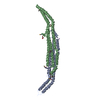
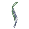
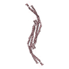
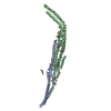
- PDB-3haj: Crystal structure of human PACSIN2 F-BAR domain (p212121 lattice) -
+ Open data

- Basic information
| Entry | Database: PDB / ID: 3haj | ||||||
|---|---|---|---|---|---|---|---|
| Title | Crystal structure of human PACSIN2 F-BAR domain (p212121 lattice) | ||||||
 Components Components | human PACSIN2 F-BAR | ||||||
 Keywords Keywords |  ENDOCYTOSIS / PACSIN / Syndapin / FAP52 / F-BAR / Alternative splicing / Coiled coil / Cytoplasmic vesicle / Phosphoprotein / Polymorphism / SH3 domain ENDOCYTOSIS / PACSIN / Syndapin / FAP52 / F-BAR / Alternative splicing / Coiled coil / Cytoplasmic vesicle / Phosphoprotein / Polymorphism / SH3 domain | ||||||
| Function / homology |  Function and homology informationcaveola assembly / cell projection morphogenesis / plasma membrane tubulation / protein localization to endosome / phosphatidic acid binding / negative regulation of endocytosis / caveolin-mediated endocytosis / regulation of endocytosis / centriolar satellite / cytoskeleton organization ...caveola assembly / cell projection morphogenesis / plasma membrane tubulation / protein localization to endosome / phosphatidic acid binding / negative regulation of endocytosis / caveolin-mediated endocytosis / regulation of endocytosis / centriolar satellite / cytoskeleton organization / cytoskeletal protein binding / caveola / modulation of chemical synaptic transmission / phospholipid binding / ruffle membrane / recycling endosome membrane / cell-cell junction / Clathrin-mediated endocytosis / actin cytoskeleton organization / early endosome / endosome / nuclear speck / cadherin binding / intracellular membrane-bounded organelle / focal adhesion / glutamatergic synapse / extracellular exosome / identical protein binding / plasma membrane / cytosol / cytoplasm Function and homology informationcaveola assembly / cell projection morphogenesis / plasma membrane tubulation / protein localization to endosome / phosphatidic acid binding / negative regulation of endocytosis / caveolin-mediated endocytosis / regulation of endocytosis / centriolar satellite / cytoskeleton organization ...caveola assembly / cell projection morphogenesis / plasma membrane tubulation / protein localization to endosome / phosphatidic acid binding / negative regulation of endocytosis / caveolin-mediated endocytosis / regulation of endocytosis / centriolar satellite / cytoskeleton organization / cytoskeletal protein binding / caveola / modulation of chemical synaptic transmission / phospholipid binding / ruffle membrane / recycling endosome membrane / cell-cell junction / Clathrin-mediated endocytosis / actin cytoskeleton organization / early endosome / endosome / nuclear speck / cadherin binding / intracellular membrane-bounded organelle / focal adhesion / glutamatergic synapse / extracellular exosome / identical protein binding / plasma membrane / cytosol / cytoplasmSimilarity search - Function | ||||||
| Biological species |  Homo sapiens (human) Homo sapiens (human) | ||||||
| Method | X-RAY DIFFRACTION / SYNCHROTRON / MOLECULAR REPLACEMENT / Resolution: 2.78 Å | ||||||
 Authors Authors | Wang, Q. / Navarro, M.V.A.S. / Peng, G. / Rajashankar, K.R. / Sondermann, H. | ||||||
 Citation Citation | Journal: Proc.Natl.Acad.Sci.USA / Year: 2009 Title: Molecular mechanism of membrane constriction and tubulation mediated by the F-BAR protein Pacsin/Syndapin. Authors: Wang, Q. / Navarro, M.V. / Peng, G. / Molinelli, E. / Lin Goh, S. / Judson, B.L. / Rajashankar, K.R. / Sondermann, H. | ||||||
| History |
|
- Structure visualization
| Structure viewer | Molecule: MolmilJmol/JSmol |
|---|
- Downloads & links
-Download
| PDBx/mmCIF format | 3haj.cif.gz | 135.3 KB | Display | PDBx/mmCIF format |
|---|---|---|---|---|
| PDB format | pdb3haj.ent.gz | 103.8 KB | Display | PDB format |
| PDBx/mmJSON format | 3haj.json.gz | Tree view | PDBx/mmJSON format | |
| Others |  Other downloads Other downloads |
-Validation report
| Arichive directory | https://data.pdbj.org/pub/pdb/validation_reports/ha/3hajftp://data.pdbj.org/pub/pdb/validation_reports/ha/3haj | HTTPS FTP |
|---|
-Related structure data
| Related structure data |  3hahC  3haiSC C: citing same article ( S: Starting model for refinement |
|---|---|
| Similar structure data |


-Links
 PDBj
PDBj
- Assembly
| Deposited unit | 
| ||||||||
|---|---|---|---|---|---|---|---|---|---|
| 1 |
| ||||||||
| Unit cell |
|
-Components
| #1: Protein | Mass: 55817.559 Da / Num. of mol.: 2 Source method: isolated from a genetically manipulated source Source: (gene. exp.) Homo sapiens (human) / Gene: PACSIN2 / Plasmid: pET28a / Production host:  Escherichia coli (E. coli) / Strain (production host): BL21(DE3) / References: UniProt: Q9UNF0 Escherichia coli (E. coli) / Strain (production host): BL21(DE3) / References: UniProt: Q9UNF0#2: Chemical |   Mass: 40.078 Da / Num. of mol.: 2 / Source method: obtained synthetically / Formula: Ca Mass: 40.078 Da / Num. of mol.: 2 / Source method: obtained synthetically / Formula: Ca#3: Water | ChemComp-HOH / | Water Mass: 18.015 Da / Num. of mol.: 36 / Source method: isolated from a natural source / Formula: H2O Mass: 18.015 Da / Num. of mol.: 36 / Source method: isolated from a natural source / Formula: H2O |
|---|
-Experimental details
-Experiment
| Experiment | Method: X-RAY DIFFRACTION |
|---|
- Sample preparation
| Crystal | Density Matthews: 2.22 Å3/Da / Density % sol: 44.5 % |
|---|---|
| Crystal grow | Temperature: 293 K / Method: vapor diffusion, hanging drop / pH: 6.5 Details: 20% PEG-MME 5000, 0.1 M Bis-Tris pH 6.5, VAPOR DIFFUSION, HANGING DROP, temperature 293K |
-Data collection
| Diffraction | Mean temperature: 110 K |
|---|---|
| Diffraction source | Source: SYNCHROTRON / Site: APS  / Beamline: 24-ID-C / Wavelength: 0.9795 Å / Beamline: 24-ID-C / Wavelength: 0.9795 Å |
| Detector | Type: ADSC QUANTUM 315 / Detector: CCD / Date: Jul 9, 2008 |
| Radiation | Protocol: SINGLE WAVELENGTH / Monochromatic (M) / Laue (L): M / Scattering type: x-ray |
| Radiation wavelength | Wavelength: 0.9795 Å / Relative weight: 1 |
| Reflection | Resolution: 2.78→44.72 Å / Num. all: 25179 / Observed criterion σ(F): 2 / Observed criterion σ(I): 2 / Redundancy: 4.2 % / Biso Wilson estimate: 78.8 Å2 / Net I/σ(I): 14.2 |
| Reflection shell | Resolution: 2.78→2.95 Å / Redundancy: 3.3 % / Mean I/σ(I) obs: 2.8 / Num. unique all: 3092 / % possible all: 30.2 |
- Processing
| Software |
| ||||||||||||||||||||||||||||||||||||||||||||||||||||||||||||||||||||||
|---|---|---|---|---|---|---|---|---|---|---|---|---|---|---|---|---|---|---|---|---|---|---|---|---|---|---|---|---|---|---|---|---|---|---|---|---|---|---|---|---|---|---|---|---|---|---|---|---|---|---|---|---|---|---|---|---|---|---|---|---|---|---|---|---|---|---|---|---|---|---|---|
| Refinement | Method to determine structure: MOLECULAR REPLACEMENT Starting model: 3HAI Resolution: 2.78→44.715 Å / SU ML: 2.2 / σ(F): 2 / Stereochemistry target values: ML
| ||||||||||||||||||||||||||||||||||||||||||||||||||||||||||||||||||||||
| Solvent computation | Shrinkage radii: 0.9 Å / VDW probe radii: 1.11 Å / Solvent model: FLAT BULK SOLVENT MODEL / Bsol: 53.3 Å2 / ksol: 0.329 e/Å3 | ||||||||||||||||||||||||||||||||||||||||||||||||||||||||||||||||||||||
| Displacement parameters | Biso mean: 78.8 Å2
| ||||||||||||||||||||||||||||||||||||||||||||||||||||||||||||||||||||||
| Refinement step | Cycle: LAST / Resolution: 2.78→44.715 Å
| ||||||||||||||||||||||||||||||||||||||||||||||||||||||||||||||||||||||
| Refine LS restraints |
| ||||||||||||||||||||||||||||||||||||||||||||||||||||||||||||||||||||||
| LS refinement shell |
|
