 Movie
Movie Controller


+ Open data

- Basic information
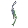
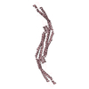
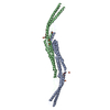
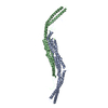
| Entry | Database: PDB / ID: 2x3v | ||||||
|---|---|---|---|---|---|---|---|
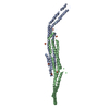
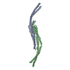
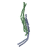
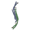
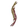
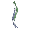
| Title | Structure of The F-BAR Domain of Mouse Syndapin I | ||||||
 Components Components | PROTEIN KINASE C AND CASEIN KINASE SUBSTRATE IN NEURONS PROTEIN 1 | ||||||
 Keywords Keywords |  ENDOCYTOSIS / BAR / N-WASP / DYNAMIN / PACSIN 1 ENDOCYTOSIS / BAR / N-WASP / DYNAMIN / PACSIN 1 | ||||||
| Function / homology |  Function and homology information Function and homology informationpresynaptic endocytic zone / plasma membrane tubulation / COPI-coated vesicle / photoreceptor ribbon synapse / Clathrin-mediated endocytosis / negative regulation of endocytosis / protein localization to membrane / regulation of postsynaptic neurotransmitter receptor internalization / positive regulation of dendrite development / regulation of endocytosis ...presynaptic endocytic zone / plasma membrane tubulation / COPI-coated vesicle / photoreceptor ribbon synapse / Clathrin-mediated endocytosis / negative regulation of endocytosis / protein localization to membrane / regulation of postsynaptic neurotransmitter receptor internalization / positive regulation of dendrite development / regulation of endocytosis / synaptic vesicle endocytosis / axon terminus / cytoskeleton organization / cytoskeletal protein binding / neuron projection morphogenesis / actin filament organization / protein localization to plasma membrane / postsynaptic density membrane / phospholipid binding / cytoplasmic vesicle membrane / ruffle membrane / myelin sheath / endosome / synapse / glutamatergic synapse / perinuclear region of cytoplasm / signal transduction / identical protein binding / plasma membrane / cytosol / cytoplasmSimilarity search - Function | ||||||
| Biological species |  MUS MUSCULUS (house mouse) MUS MUSCULUS (house mouse) | ||||||
| Method | X-RAY DIFFRACTION / SYNCHROTRON / MAD / Resolution: 2.45 Å | ||||||
 Authors Authors | Ma, Q. / Rao, Y. / Vahedi-Faridi, A. / Saenger, W. / Haucke, V. | ||||||
 Citation Citation | Journal: Proc.Natl.Acad.Sci.USA / Year: 2010 Title: Molecular Basis for SH3 Domain Regulation of F-Bar-Mediated Membrane Deformation. Authors: Rao, Y. / Ma, Q. / Vahedi-Faridi, A. / Sundborger, A. / Pechstein, A. / Puchkov, D. / Luo, L. / Shupliakov, O. / Saenger, W. / Haucke, V. | ||||||
| History |
|
- Structure visualization
| Structure viewer | Molecule: MolmilJmol/JSmol |
|---|
- Downloads & links
-Download
| PDBx/mmCIF format | 2x3v.cif.gz | 188.6 KB | Display | PDBx/mmCIF format |
|---|---|---|---|---|
| PDB format | pdb2x3v.ent.gz | 154.2 KB | Display | PDB format |
| PDBx/mmJSON format | 2x3v.json.gz | Tree view | PDBx/mmJSON format | |
| Others |  Other downloads Other downloads |
-Validation report
| Arichive directory | https://data.pdbj.org/pub/pdb/validation_reports/x3/2x3vftp://data.pdbj.org/pub/pdb/validation_reports/x3/2x3v | HTTPS FTP |
|---|
-Related structure data


-Links
 PDBj
PDBj


- Assembly
| Deposited unit | 
| ||||||||
|---|---|---|---|---|---|---|---|---|---|
| 1 | 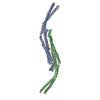
| ||||||||
| 2 | 
| ||||||||
| Unit cell |
|
-Components
| #1: Protein | Mass: 39349.430 Da / Num. of mol.: 3 / Fragment: F-BAR DOMAIN, RESIDUES 1-337 Source method: isolated from a genetically manipulated source Source: (gene. exp.) MUS MUSCULUS (house mouse) / Plasmid: PET28A / Production host:  ESCHERICHIA COLI (E. coli) / Strain (production host): BL21(DE3) / References: UniProt: Q61644 ESCHERICHIA COLI (E. coli) / Strain (production host): BL21(DE3) / References: UniProt: Q61644#2: Water | ChemComp-HOH / | Water Mass: 18.015 Da / Num. of mol.: 158 / Source method: isolated from a natural source / Formula: H2O Mass: 18.015 Da / Num. of mol.: 158 / Source method: isolated from a natural source / Formula: H2OSequence details | THE PROTEIN CRYSTALLIZ | |
|---|
-Experimental details
-Experiment
| Experiment | Method: X-RAY DIFFRACTION / Number of used crystals: 1 |
|---|
- Sample preparation
| Crystal | Density Matthews: 3.1 Å3/Da / Density % sol: 60 % / Description: NONE |
|---|---|
| Crystal grow | pH: 5.5 Details: 2UL SYNDAPIN-I/EHD-1(401 -534)(25MG/ML IN 50MM HEPES,200MM NACL) MIXED WITH WELL SOLUTION 0.1M SODIUM CITRATE BUFFER (PH5.5),0.2M NAAC,10%(W/V) PEG4000. ONLY F-BAR DOMAIN OF SYDAPIN I CRYSTALLIZED. |
-Data collection
| Diffraction | Mean temperature: 100 K |
|---|---|
| Diffraction source | Source: SYNCHROTRON / Site: BESSY  / Beamline: 14.1 / Wavelength: 0.978 / Beamline: 14.1 / Wavelength: 0.978 |
| Detector | Type: MARMOSAIC 225 mm CCD / Detector: CCD / Date: Aug 15, 2009 Details: DOUBLE CRYSTAL MONOCHROMATOR WITH 2 SETS OF MIRRORS |
| Radiation | Monochromator: DOUBLE CRYSTAL / Protocol: MAD / Monochromatic (M) / Laue (L): M / Scattering type: x-ray |
| Radiation wavelength | Wavelength: 0.978 Å / Relative weight: 1 |
| Reflection | Resolution: 2.45→76.75 Å / Num. obs: 50261 / % possible obs: 97.2 % / Observed criterion σ(I): -3 / Redundancy: 6.69 % / Biso Wilson estimate: 55.7 Å2 / Rmerge(I) obs: 0.08 / Net I/σ(I): 16.35 |
| Reflection shell | Resolution: 2.45→2.55 Å / Redundancy: 3.96 % / Rmerge(I) obs: 0.84 / Mean I/σ(I) obs: 1.84 / % possible all: 81.8 |
- Processing
| Software |
| ||||||||||||||||||||||||||||||||||||||||||||||||||||||||||||||||||||||||||||||||||||||||||||||||||||||||||||||||||||||||||||||||||||||||||||||||||||||||||||||||||||||||||||||||||||||
|---|---|---|---|---|---|---|---|---|---|---|---|---|---|---|---|---|---|---|---|---|---|---|---|---|---|---|---|---|---|---|---|---|---|---|---|---|---|---|---|---|---|---|---|---|---|---|---|---|---|---|---|---|---|---|---|---|---|---|---|---|---|---|---|---|---|---|---|---|---|---|---|---|---|---|---|---|---|---|---|---|---|---|---|---|---|---|---|---|---|---|---|---|---|---|---|---|---|---|---|---|---|---|---|---|---|---|---|---|---|---|---|---|---|---|---|---|---|---|---|---|---|---|---|---|---|---|---|---|---|---|---|---|---|---|---|---|---|---|---|---|---|---|---|---|---|---|---|---|---|---|---|---|---|---|---|---|---|---|---|---|---|---|---|---|---|---|---|---|---|---|---|---|---|---|---|---|---|---|---|---|---|---|---|
| Refinement | Method to determine structure: MAD Starting model: NONE Resolution: 2.45→76.7 Å / Cor.coef. Fo:Fc: 0.952 / Cor.coef. Fo:Fc free: 0.934 / SU B: 15.91 / SU ML: 0.168 / TLS residual ADP flag: LIKELY RESIDUAL / Cross valid method: THROUGHOUT / ESU R: 0.337 / ESU R Free: 0.255 / Stereochemistry target values: MAXIMUM LIKELIHOOD / Details: HYDROGENS HAVE BEEN ADDED IN THE RIDING POSITIONS.
| ||||||||||||||||||||||||||||||||||||||||||||||||||||||||||||||||||||||||||||||||||||||||||||||||||||||||||||||||||||||||||||||||||||||||||||||||||||||||||||||||||||||||||||||||||||||
| Solvent computation | Ion probe radii: 0.8 Å / Shrinkage radii: 0.8 Å / VDW probe radii: 1.2 Å / Solvent model: MASK | ||||||||||||||||||||||||||||||||||||||||||||||||||||||||||||||||||||||||||||||||||||||||||||||||||||||||||||||||||||||||||||||||||||||||||||||||||||||||||||||||||||||||||||||||||||||
| Displacement parameters | Biso mean: 32.69 Å2
| ||||||||||||||||||||||||||||||||||||||||||||||||||||||||||||||||||||||||||||||||||||||||||||||||||||||||||||||||||||||||||||||||||||||||||||||||||||||||||||||||||||||||||||||||||||||
| Refinement step | Cycle: LAST / Resolution: 2.45→76.7 Å
| ||||||||||||||||||||||||||||||||||||||||||||||||||||||||||||||||||||||||||||||||||||||||||||||||||||||||||||||||||||||||||||||||||||||||||||||||||||||||||||||||||||||||||||||||||||||
| Refine LS restraints |
|