 Movie
Movie Controller

[English] 日本語
 Yorodumi
- PDB-3h6e: The crystal structure of a carbohydrate kinase from Novosphingobi... -
+ Open data

- Basic information
| Entry | Database: PDB / ID: 3h6e | ||||||
|---|---|---|---|---|---|---|---|
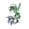
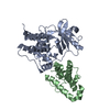
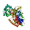
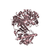
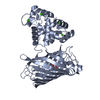
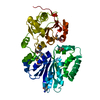
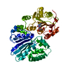
| Title | The crystal structure of a carbohydrate kinase from Novosphingobium aromaticivorans | ||||||
 Components Components | Carbohydrate kinase, FGGY | ||||||
 Keywords Keywords |  TRANSFERASE / carbohydrate kinase / Novosphingobium aromaticivorans / strain DSM 12444 / SGX / 11200i / Kinase / Structural Genomics / PSI-2 / Protein Structure Initiative / New York SGX Research Center for Structural Genomics / NYSGXRC TRANSFERASE / carbohydrate kinase / Novosphingobium aromaticivorans / strain DSM 12444 / SGX / 11200i / Kinase / Structural Genomics / PSI-2 / Protein Structure Initiative / New York SGX Research Center for Structural Genomics / NYSGXRC | ||||||
| Function / homology |  Function and homology information Function and homology informationphosphotransferase activity, alcohol group as acceptor / kinase activity / carbohydrate metabolic processSimilarity search - Function | ||||||
| Biological species |  Novosphingobium aromaticivorans (bacteria) Novosphingobium aromaticivorans (bacteria) | ||||||
| Method | X-RAY DIFFRACTION / SYNCHROTRON / SAD / Resolution: 2.5 Å | ||||||
 Authors Authors | Zhang, Z. / Burley, S.K. / Swaminathan, S. / New York SGX Research Center for Structural Genomics (NYSGXRC) | ||||||
 Citation Citation | Journal: To be Published Title: The crystal structure of carbohydrate kinase from Novosphingobium aromaticivorans Authors: Zhang, Z. / Burley, S.K. / Swaminathan, S. | ||||||
| History |
|
- Structure visualization
| Structure viewer | Molecule: MolmilJmol/JSmol |
|---|
- Downloads & links
-Download
| PDBx/mmCIF format | 3h6e.cif.gz | 188.7 KB | Display | PDBx/mmCIF format |
|---|---|---|---|---|
| PDB format | pdb3h6e.ent.gz | 157.1 KB | Display | PDB format |
| PDBx/mmJSON format | 3h6e.json.gz | Tree view | PDBx/mmJSON format | |
| Others |  Other downloads Other downloads |
-Validation report
| Arichive directory | https://data.pdbj.org/pub/pdb/validation_reports/h6/3h6eftp://data.pdbj.org/pub/pdb/validation_reports/h6/3h6e | HTTPS FTP |
|---|
-Related structure data
| Similar structure data | |
|---|---|
| Other databases |


-Links
 PDBj
PDBj- Assembly
| Deposited unit | 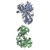
| ||||||||
|---|---|---|---|---|---|---|---|---|---|
| 1 | 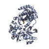
| ||||||||
| 2 | 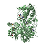
| ||||||||
| Unit cell |
|
-Components
| #1: Protein | Mass: 53312.941 Da / Num. of mol.: 2 Source method: isolated from a genetically manipulated source Source: (gene. exp.) Novosphingobium aromaticivorans (bacteria)Strain: DSM 12444 / Gene: Saro_1051 / Plasmid: BC-pSGX3(BC) / Production host: Escherichia coli BL21(DE3) (bacteria) / Strain (production host): BL21(DE3)-codon+RIL - Stratagene / References: UniProt: Q2G9H7#2: Water | ChemComp-HOH / | Water Mass: 18.015 Da / Num. of mol.: 321 / Source method: isolated from a natural source / Formula: H2O Mass: 18.015 Da / Num. of mol.: 321 / Source method: isolated from a natural source / Formula: H2O |
|---|
-Experimental details
-Experiment
| Experiment | Method: X-RAY DIFFRACTION / Number of used crystals: 1 |
|---|
- Sample preparation
| Crystal | Density Matthews: 2.3 Å3/Da / Density % sol: 46.41 % |
|---|---|
| Crystal grow | Temperature: 291 K / Method: vapor diffusion, sitting drop / pH: 6.5 Details: 0.2 M Magnesesium chloride hexahydrate, 0.1 M BIS-TRIS pH 6.5, 25% w/v PEG 3350, VAPOR DIFFUSION, SITTING DROP, temperature 291K |
-Data collection
| Diffraction | Mean temperature: 100 K |
|---|---|
| Diffraction source | Source: SYNCHROTRON / Site: NSLS  / Beamline: X29A / Wavelength: 0.9792 Å / Beamline: X29A / Wavelength: 0.9792 Å |
| Detector | Type: ADSC QUANTUM 315 / Detector: CCD / Date: Apr 12, 2009 / Details: mirrors |
| Radiation | Monochromator: Si 111 CHANNEL / Protocol: SINGLE WAVELENGTH / Monochromatic (M) / Laue (L): M / Scattering type: x-ray |
| Radiation wavelength | Wavelength: 0.9792 Å / Relative weight: 1 |
| Reflection | Resolution: 2.5→114.71 Å / Num. obs: 35006 / % possible obs: 100 % / Observed criterion σ(F): 0 / Observed criterion σ(I): 0 / Redundancy: 13.7 % / Biso Wilson estimate: 38.41 Å2 / Rmerge(I) obs: 0.082 / Net I/σ(I): 29.3 |
| Reflection shell | Resolution: 2.5→2.64 Å / Redundancy: 13.8 % / Rmerge(I) obs: 0.191 / Mean I/σ(I) obs: 11.5 / % possible all: 99.9 |
- Processing
| Software |
| ||||||||||||||||||||||||||||||||||||||||||||||||||||||||||||||||||||||||||||||||||||||||||||||||||
|---|---|---|---|---|---|---|---|---|---|---|---|---|---|---|---|---|---|---|---|---|---|---|---|---|---|---|---|---|---|---|---|---|---|---|---|---|---|---|---|---|---|---|---|---|---|---|---|---|---|---|---|---|---|---|---|---|---|---|---|---|---|---|---|---|---|---|---|---|---|---|---|---|---|---|---|---|---|---|---|---|---|---|---|---|---|---|---|---|---|---|---|---|---|---|---|---|---|---|---|
| Refinement | Method to determine structure: SAD / Resolution: 2.5→51.279 Å / SU ML: 0.37 / Isotropic thermal model: isotropic / σ(F): 1.14 / Phase error: 26.25 / Stereochemistry target values: ML
| ||||||||||||||||||||||||||||||||||||||||||||||||||||||||||||||||||||||||||||||||||||||||||||||||||
| Solvent computation | Shrinkage radii: 0.9 Å / VDW probe radii: 1.11 Å / Solvent model: FLAT BULK SOLVENT MODEL / Bsol: 36.755 Å2 / ksol: 0.355 e/Å3 | ||||||||||||||||||||||||||||||||||||||||||||||||||||||||||||||||||||||||||||||||||||||||||||||||||
| Displacement parameters | Biso mean: 27.29 Å2
| ||||||||||||||||||||||||||||||||||||||||||||||||||||||||||||||||||||||||||||||||||||||||||||||||||
| Refinement step | Cycle: LAST / Resolution: 2.5→51.279 Å
| ||||||||||||||||||||||||||||||||||||||||||||||||||||||||||||||||||||||||||||||||||||||||||||||||||
| Refine LS restraints |
| ||||||||||||||||||||||||||||||||||||||||||||||||||||||||||||||||||||||||||||||||||||||||||||||||||
| LS refinement shell |
|
