 Movie
Movie Controller


+ Open data

- Basic information
| Entry | Database: PDB / ID: 2c83 | ||||||
|---|---|---|---|---|---|---|---|
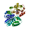
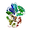
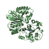
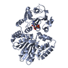
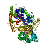
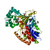
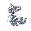
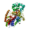
| Title | CRYSTAL STRUCTURE OF THE SIALYLTRANSFERASE PM0188 | ||||||
 Components Components | HYPOTHETICAL PROTEIN PM0188 Hypothesis Hypothesis | ||||||
 Keywords Keywords | TRANSFERASE / HYPOTHETICAL PROTEIN / PM0188 / SIALYLTRANSFERASE | ||||||
| Function / homology |  Function and homology information Function and homology information | ||||||
| Biological species |  PASTEURELLA MULTOCIDA (bacteria) PASTEURELLA MULTOCIDA (bacteria) | ||||||
| Method | X-RAY DIFFRACTION / SYNCHROTRON / MAD / Resolution: 1.9 Å | ||||||
 Authors Authors | Kim, D.U. / Cho, H.S. | ||||||
 Citation Citation | Journal: Bmb Rep / Year: 2008 Title: Structural analysis of sialyltransferase PM0188 from Pasteurella multocida complexed with donor analogue and acceptor sugar. Authors: Kim, D.U. / Yoo, J.H. / Lee, Y.J. / Kim, K.S. / Cho, H.S. #1: Journal: Nat.Struct.Mol.Biol. / Year: 2004Title: Structural Analysis of the Sialyltransferase Cstii from Campylobacter Jejuni in Complex with a Substrate Analog Authors: Chiu, C.P. / Watts, A.G. / Lairson, L.L. / Gilbert, M. / Lim, D. / Wakarchuk, W.W. / Withers, S.G. / Strynadka, N.C. | ||||||
| History |
|
- Structure visualization
| Structure viewer | Molecule: MolmilJmol/JSmol |
|---|
- Downloads & links
-Download
| PDBx/mmCIF format | 2c83.cif.gz | 90.2 KB | Display | PDBx/mmCIF format |
|---|---|---|---|---|
| PDB format | pdb2c83.ent.gz | 72.5 KB | Display | PDB format |
| PDBx/mmJSON format | 2c83.json.gz | Tree view | PDBx/mmJSON format | |
| Others |  Other downloads Other downloads |
-Validation report
| Arichive directory | https://data.pdbj.org/pub/pdb/validation_reports/c8/2c83ftp://data.pdbj.org/pub/pdb/validation_reports/c8/2c83 | HTTPS FTP |
|---|
-Related structure data


-Links
 PDBj
PDBj- Assembly
| Deposited unit | 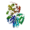
| ||||||||
|---|---|---|---|---|---|---|---|---|---|
| 1 |
| ||||||||
| Unit cell |
|
-Components
| #1: Protein | Hypothesis / ALPHA-2 / 6-SIALYLTRANSFERASE PM0188 Mass: 45689.582 Da / Num. of mol.: 1 / Fragment: RESIDUES 26-412 Source method: isolated from a genetically manipulated source Source: (gene. exp.) PASTEURELLA MULTOCIDA (bacteria) / Production host: ESCHERICHIA COLI (E. coli) / Strain (production host): BL21(DE3) / References: UniProt: Q9CP67, UniProt: Q15KI8*PLUS |
|---|---|
| #2: Water | ChemComp-HOH / Water Mass: 18.015 Da / Num. of mol.: 132 / Source method: isolated from a natural source / Formula: H2O Mass: 18.015 Da / Num. of mol.: 132 / Source method: isolated from a natural source / Formula: H2O |
-Experimental details
-Experiment
| Experiment | Method: X-RAY DIFFRACTION / Number of used crystals: 1 |
|---|
- Sample preparation
| Crystal | Density Matthews: 2.1 Å3/Da / Density % sol: 40.8 % / Description: NONE |
|---|---|
| Crystal grow | pH: 6.5 / Details: pH 6.5 |
-Data collection
| Diffraction | Mean temperature: 100 K | |||||||||
|---|---|---|---|---|---|---|---|---|---|---|
| Diffraction source | Source: SYNCHROTRON / Site: PAL/PLS  / Beamline: 6B / Wavelength: 0.9713700, 0.9790300 / Beamline: 6B / Wavelength: 0.9713700, 0.9790300 | |||||||||
| Detector | Type: BRUCKER / Detector: CCD | |||||||||
| Radiation | Protocol: MAD / Monochromatic (M) / Laue (L): M / Scattering type: x-ray | |||||||||
| Radiation wavelength |
| |||||||||
| Reflection | Resolution: 1.9→30 Å / Num. obs: 30006 / % possible obs: 98.7 % / Observed criterion σ(I): 1 / Redundancy: 5.3 % / Rmerge(I) obs: 0.11 / Net I/σ(I): 15.4 | |||||||||
| Reflection shell | Resolution: 1.9→1.97 Å / Redundancy: 3.3 % / Rmerge(I) obs: 1.4 / Mean I/σ(I) obs: 2.1 / % possible all: 93.1 |
- Processing
| Software |
| ||||||||||||||||||||||||||||||||||||||||||||||||||||||||||||
|---|---|---|---|---|---|---|---|---|---|---|---|---|---|---|---|---|---|---|---|---|---|---|---|---|---|---|---|---|---|---|---|---|---|---|---|---|---|---|---|---|---|---|---|---|---|---|---|---|---|---|---|---|---|---|---|---|---|---|---|---|---|
| Refinement | Method to determine structure: MAD Starting model: NONE Resolution: 1.9→15 Å / Data cutoff high absF: 10000 / Cross valid method: THROUGHOUT / σ(F): 0
| ||||||||||||||||||||||||||||||||||||||||||||||||||||||||||||
| Solvent computation | Bsol: 41.8875 Å2 / ksol: 0.356479 e/Å3 | ||||||||||||||||||||||||||||||||||||||||||||||||||||||||||||
| Displacement parameters |
| ||||||||||||||||||||||||||||||||||||||||||||||||||||||||||||
| Refinement step | Cycle: LAST / Resolution: 1.9→15 Å
| ||||||||||||||||||||||||||||||||||||||||||||||||||||||||||||
| Refine LS restraints |
| ||||||||||||||||||||||||||||||||||||||||||||||||||||||||||||
| Xplor file |
|
