 Movie
Movie Controller

[English] 日本語
 Yorodumi
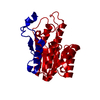
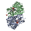
- PDB-3guy: Crystal structure of a short-chain dehydrogenase/reductase from V... -
+ Open data

- Basic information
| Entry | Database: PDB / ID: 3guy | ||||||
|---|---|---|---|---|---|---|---|
| Title | Crystal structure of a short-chain dehydrogenase/reductase from Vibrio parahaemolyticus | ||||||
 Components Components | Short-chain dehydrogenase/reductase SDR | ||||||
 Keywords Keywords |  OXIDOREDUCTASE / STRUCTURAL GENOMICS / PSI-2 / Protein Structure Initiative / New York SGX Research Center for Structural Genomics / NYSGXRC OXIDOREDUCTASE / STRUCTURAL GENOMICS / PSI-2 / Protein Structure Initiative / New York SGX Research Center for Structural Genomics / NYSGXRC | ||||||
| Function / homology | short chain dehydrogenase / Short-chain dehydrogenase/reductase SDR / NAD(P)-binding Rossmann-like Domain / NAD(P)-binding domain superfamily / Rossmann fold / 3-Layer(aba) Sandwich / Alpha Beta / Short-chain dehydrogenase/reductase SDR Function and homology information Function and homology information | ||||||
| Biological species |   Vibrio parahaemolyticus (bacteria) Vibrio parahaemolyticus (bacteria) | ||||||
| Method | X-RAY DIFFRACTION / SYNCHROTRON / SAD / Resolution: 1.9 Å | ||||||
 Authors Authors | Patskovsky, Y. / Bonanno, J.B. / Freeman, J. / Bain, K.T. / Miller, S. / Sampathkumar, P. / Wasserman, S. / Sauder, J.M. / Burley, S.K. / Almo, S.C. / New York SGX Research Center for Structural Genomics (NYSGXRC) | ||||||
 Citation Citation | Journal: To be Published Title: Crystal structure of a short-chain dehydrogenase/reductase from Vibrio parahaemolyticus Authors: Patskovsky, Y. / Bonanno, J.B. / Freeman, J. / Bain, K.T. / Miller, S. / Sampathkumar, P. / Wasserman, S. / Sauder, J.M. / Burley, S.K. / Almo, S.C. | ||||||
| History |
|
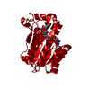
- Structure visualization
| Structure viewer | Molecule: MolmilJmol/JSmol |
|---|
- Downloads & links
-Download
| PDBx/mmCIF format | 3guy.cif.gz | 327.5 KB | Display | PDBx/mmCIF format |
|---|---|---|---|---|
| PDB format | pdb3guy.ent.gz | 266.3 KB | Display | PDB format |
| PDBx/mmJSON format | 3guy.json.gz | Tree view | PDBx/mmJSON format | |
| Others |  Other downloads Other downloads |
-Validation report
| Arichive directory | https://data.pdbj.org/pub/pdb/validation_reports/gu/3guyftp://data.pdbj.org/pub/pdb/validation_reports/gu/3guy | HTTPS FTP |
|---|
-Related structure data
| Similar structure data | |
|---|---|
| Other databases |


-Links
 PDBj
PDBj

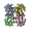
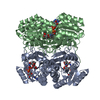
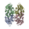
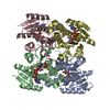
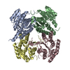
- Assembly
| Deposited unit | 
| ||||||||
|---|---|---|---|---|---|---|---|---|---|
| 1 | 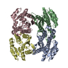
| ||||||||
| 2 | 
| ||||||||
| Unit cell |
| ||||||||
| Details | probable tetramer |
-Components
| #1: Protein | Mass: 25034.264 Da / Num. of mol.: 8 / Mutation: C52R, S204A Source method: isolated from a genetically manipulated source Source: (gene. exp.) Vibrio parahaemolyticus (bacteria) / Strain: AQ3810 / Gene: A79_5576 / Plasmid: modified pET26 / Production host: Escherichia coli (E. coli) / Strain (production host): BL21(DE3) / References: UniProt: A6B7Q2#2: Water | ChemComp-HOH / | Water Mass: 18.015 Da / Num. of mol.: 586 / Source method: isolated from a natural source / Formula: H2O Mass: 18.015 Da / Num. of mol.: 586 / Source method: isolated from a natural source / Formula: H2O |
|---|
-Experimental details
-Experiment
| Experiment | Method: X-RAY DIFFRACTION / Number of used crystals: 1 |
|---|
- Sample preparation
| Crystal | Density Matthews: 2.09 Å3/Da / Density % sol: 41.04 % |
|---|---|
| Crystal grow | Temperature: 294 K / Method: vapor diffusion / pH: 5.4 Details: 100mM Bis-Tris pH 5.4, 15% PEG 3350, 100mM magnesium chloride, vapor diffusion, temperature 294K |
-Data collection
| Diffraction | Mean temperature: 100 K |
|---|---|
| Diffraction source | Source: SYNCHROTRON / Site: APS  / Beamline: 31-ID / Wavelength: 0.97958 Å / Beamline: 31-ID / Wavelength: 0.97958 Å |
| Detector | Type: MAR CCD 165 mm / Detector: CCD / Date: Feb 3, 2009 |
| Radiation | Monochromator: diamond / Protocol: SINGLE WAVELENGTH / Monochromatic (M) / Laue (L): M / Scattering type: x-ray |
| Radiation wavelength | Wavelength: 0.97958 Å / Relative weight: 1 |
| Reflection | Resolution: 1.9→88.44 Å / Num. all: 127600 / Num. obs: 123644 / % possible obs: 96.9 % / Observed criterion σ(F): 0 / Observed criterion σ(I): 0 / Redundancy: 2.4 % / Biso Wilson estimate: 23.7 Å2 / Rmerge(I) obs: 0.089 / Rsym value: 0.089 / Net I/σ(I): 7.3 |
| Reflection shell | Resolution: 1.9→2 Å / Redundancy: 2.4 % / Rmerge(I) obs: 0.324 / Mean I/σ(I) obs: 2.1 / Num. measured all: 43304 / Num. unique all: 17750 / Rsym value: 0.324 / % possible all: 95.5 |
- Processing
| Software |
| |||||||||||||||||||||||||||||||||||||||||||||||||||||||||||||||||||||||||||||||||||||
|---|---|---|---|---|---|---|---|---|---|---|---|---|---|---|---|---|---|---|---|---|---|---|---|---|---|---|---|---|---|---|---|---|---|---|---|---|---|---|---|---|---|---|---|---|---|---|---|---|---|---|---|---|---|---|---|---|---|---|---|---|---|---|---|---|---|---|---|---|---|---|---|---|---|---|---|---|---|---|---|---|---|---|---|---|---|---|
| Refinement | Method to determine structure: SAD / Resolution: 1.9→20 Å / Cor.coef. Fo:Fc: 0.953 / Cor.coef. Fo:Fc free: 0.926 / WRfactor Rfree: 3.425 / WRfactor Rwork: 2.225 / Occupancy max: 1 / Occupancy min: 1 / FOM work R set: 0.811 / SU B: 4.189 / SU ML: 0.121 / SU R Cruickshank DPI: 0.178 / SU Rfree: 0.164 / Cross valid method: THROUGHOUT / σ(F): 0 / σ(I): 0 / ESU R: 0.178 / ESU R Free: 0.164 / Stereochemistry target values: MAXIMUM LIKELIHOOD / Details: HYDROGENS HAVE BEEN ADDED IN THE RIDING POSITIONS
| |||||||||||||||||||||||||||||||||||||||||||||||||||||||||||||||||||||||||||||||||||||
| Solvent computation | Ion probe radii: 0.8 Å / Shrinkage radii: 0.8 Å / VDW probe radii: 1.2 Å / Solvent model: BABINET MODEL WITH MASK | |||||||||||||||||||||||||||||||||||||||||||||||||||||||||||||||||||||||||||||||||||||
| Displacement parameters | Biso max: 66.59 Å2 / Biso mean: 32.568 Å2 / Biso min: 9.28 Å2
| |||||||||||||||||||||||||||||||||||||||||||||||||||||||||||||||||||||||||||||||||||||
| Refinement step | Cycle: LAST / Resolution: 1.9→20 Å
| |||||||||||||||||||||||||||||||||||||||||||||||||||||||||||||||||||||||||||||||||||||
| Refine LS restraints |
| |||||||||||||||||||||||||||||||||||||||||||||||||||||||||||||||||||||||||||||||||||||
| LS refinement shell | Resolution: 1.9→1.949 Å / Total num. of bins used: 20
|
