 Movie
Movie Controller

[English] 日本語
 Yorodumi
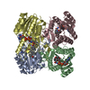
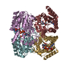
- PDB-2ewm: Crystal Structure of the (S)-Specific 1-Phenylethanol Dehydrogena... -
+ Open data

- Basic information
| Entry | Database: PDB / ID: 2ewm | ||||||
|---|---|---|---|---|---|---|---|
| Title | Crystal Structure of the (S)-Specific 1-Phenylethanol Dehydrogenase of the Denitrifying Bacterium Strain EbN1 | ||||||
 Components Components | (S)-1-Phenylethanol dehydrogenase | ||||||
 Keywords Keywords |  TRANSFERASE / Dehydrogenase TRANSFERASE / Dehydrogenase | ||||||
| Function / homology |  Function and homology information(S)-1-phenylethanol dehydrogenase / anaerobic ethylbenzene catabolic process / oxidoreductase activity, acting on the CH-OH group of donors, NAD or NADP as acceptor / protein homotetramerization / nucleotide binding Function and homology information(S)-1-phenylethanol dehydrogenase / anaerobic ethylbenzene catabolic process / oxidoreductase activity, acting on the CH-OH group of donors, NAD or NADP as acceptor / protein homotetramerization / nucleotide bindingSimilarity search - Function | ||||||
| Biological species |  Azoarcus (bacteria) Azoarcus (bacteria) | ||||||
| Method | X-RAY DIFFRACTION / SYNCHROTRON / MOLECULAR REPLACEMENT / Resolution: 2.4 Å | ||||||
 Authors Authors | Hoeffken, H.W. | ||||||
 Citation Citation | Journal: Biochemistry / Year: 2006 Title: Crystal Structure and Enzyme kinetics of the (S)-Specific 1-Phenylethanol Dehydrogenase of the denitrifying bacterium Strain EbN1 Authors: Hoeffken, H.W. / Duong, M. / Friedrich, T. / Breuer, M. / Hauer, B. / Reinhardt, R. / Rabus, R. / Heider, J. | ||||||
| History |
|
- Structure visualization
| Structure viewer | Molecule: MolmilJmol/JSmol |
|---|
- Downloads & links
-Download
| PDBx/mmCIF format | 2ewm.cif.gz | 108.1 KB | Display | PDBx/mmCIF format |
|---|---|---|---|---|
| PDB format | pdb2ewm.ent.gz | 83.9 KB | Display | PDB format |
| PDBx/mmJSON format | 2ewm.json.gz | Tree view | PDBx/mmJSON format | |
| Others |  Other downloads Other downloads |
-Validation report
| Arichive directory | https://data.pdbj.org/pub/pdb/validation_reports/ew/2ewmftp://data.pdbj.org/pub/pdb/validation_reports/ew/2ewm | HTTPS FTP |
|---|
-Related structure data
| Related structure data |  2ew8SC S: Starting model for refinement C: citing same article ( |
|---|---|
| Similar structure data |


-Links
 PDBj
PDBj

- Assembly
| Deposited unit | 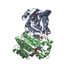
| ||||||||||||
|---|---|---|---|---|---|---|---|---|---|---|---|---|---|
| 1 | 
| ||||||||||||
| Unit cell |
| ||||||||||||
| Components on special symmetry positions |
| ||||||||||||
| Details | the biological assembly is the tetramer generated by a two fold axis |
-Components
| #1: Protein | Mass: 26687.668 Da / Num. of mol.: 2 Source method: isolated from a genetically manipulated source Source: (gene. exp.) Azoarcus (bacteria) / Genus: Azoarcus / Production host: Escherichia coli (E. coli) / Strain (production host): TG1 / References: GenBank: 56476740, UniProt: Q5P5I4*PLUS#2: Chemical | Sulfate  Mass: 96.063 Da / Num. of mol.: 2 / Source method: obtained synthetically / Formula: SO4 Mass: 96.063 Da / Num. of mol.: 2 / Source method: obtained synthetically / Formula: SO4#3: Chemical | Nicotinamide adenine dinucleotide  Mass: 663.425 Da / Num. of mol.: 2 / Source method: obtained synthetically / Formula: C21H27N7O14P2 / Comment: NAD*YM Mass: 663.425 Da / Num. of mol.: 2 / Source method: obtained synthetically / Formula: C21H27N7O14P2 / Comment: NAD*YM#4: Water | ChemComp-HOH / | Water Mass: 18.015 Da / Num. of mol.: 167 / Source method: isolated from a natural source / Formula: H2O Mass: 18.015 Da / Num. of mol.: 167 / Source method: isolated from a natural source / Formula: H2O |
|---|
-Experimental details
-Experiment
| Experiment | Method: X-RAY DIFFRACTION / Number of used crystals: 1 |
|---|
- Sample preparation
| Crystal | Density Matthews: 1.89 Å3/Da / Density % sol: 34.78 % |
|---|---|
| Crystal grow | Temperature: 294 K / Method: vapor diffusion, sitting drop / pH: 7.5 Details: 12-18% PEG 8000, 0.05M potassium phasphate, 3% 2-methyl-2,4-pentanediol crystal was soaked with 5mM NAD+ and 5mM (R)-1-phenylethanol, pH 7.5, VAPOR DIFFUSION, SITTING DROP, temperature 294K |
-Data collection
| Diffraction | Mean temperature: 80 K |
|---|---|
| Diffraction source | Source: SYNCHROTRON / Site: SLS  / Beamline: X06SA / Wavelength: 1.00885 Å / Beamline: X06SA / Wavelength: 1.00885 Å |
| Detector | Type: PSI PILATUS 6M / Detector: PIXEL / Date: Jan 31, 2005 |
| Radiation | Protocol: SINGLE WAVELENGTH / Monochromatic (M) / Laue (L): M / Scattering type: x-ray |
| Radiation wavelength | Wavelength: 1.00885 Å / Relative weight: 1 |
| Reflection | Resolution: 2.4→28 Å / Num. obs: 16156 / % possible obs: 92 % / Observed criterion σ(F): 0 / Observed criterion σ(I): 0 / Rmerge(I) obs: 0.073 / Rsym value: 0.08 / Net I/σ(I): 14.5 |
| Reflection shell | Resolution: 2.4→2.5 Å / Rmerge(I) obs: 0.122 / Num. unique all: 3088 / % possible all: 86.8 |
- Processing
| Software |
| |||||||||||||||||||||||||
|---|---|---|---|---|---|---|---|---|---|---|---|---|---|---|---|---|---|---|---|---|---|---|---|---|---|---|
| Refinement | Method to determine structure: MOLECULAR REPLACEMENT Starting model: PDB ENTRY 2EW8 Resolution: 2.4→28 Å / σ(F): 0 / Stereochemistry target values: Engh & Huber
| |||||||||||||||||||||||||
| Refine analyze |
| |||||||||||||||||||||||||
| Refinement step | Cycle: LAST / Resolution: 2.4→28 Å
| |||||||||||||||||||||||||
| Refine LS restraints |
| |||||||||||||||||||||||||
| LS refinement shell | Resolution: 2.4→2.55 Å / Rfactor Rfree error: 0.026
|
