 Movie
Movie Controller

[English] 日本語
 Yorodumi
- PDB-3gpd: TWINNING IN CRYSTALS OF HUMAN SKELETAL MUSCLE D-GLYCERALDEHYDE-3-... -
+ Open data

- Basic information
| Entry | Database: PDB / ID: 3gpd | ||||||
|---|---|---|---|---|---|---|---|
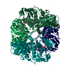
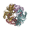
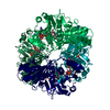
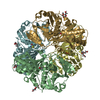
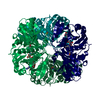
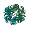
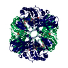
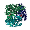
| Title | TWINNING IN CRYSTALS OF HUMAN SKELETAL MUSCLE D-GLYCERALDEHYDE-3-PHOSPHATE DEHYDROGENASE | ||||||
 Components Components | D-GLYCERALDEHYDE-3-PHOSPHATE DEHYDROGENASE | ||||||
 Keywords Keywords | OXIDOREDUCTASE (NAD(A)-ALDEHYDE(D)) | ||||||
| Function / homology |  Function and homology information Function and homology informationpositive regulation of killing of cells of another organism => GO:0051712 / positive regulation of cytokine production => GO:0001819 / peptidyl-cysteine S-trans-nitrosylation /  Transferases; Transferring nitrogenous groups; Transferring other nitrogenous groups / peptidyl-cysteine S-nitrosylase activity / killing by host of symbiont cells / glyceraldehyde-3-phosphate dehydrogenase (phosphorylating) / negative regulation of endopeptidase activity / glyceraldehyde-3-phosphate dehydrogenase (NAD+) (phosphorylating) activity / aspartic-type endopeptidase inhibitor activity ...positive regulation of killing of cells of another organism => GO:0051712 / positive regulation of cytokine production => GO:0001819 / peptidyl-cysteine S-trans-nitrosylation / Transferases; Transferring nitrogenous groups; Transferring other nitrogenous groups / peptidyl-cysteine S-nitrosylase activity / killing by host of symbiont cells / glyceraldehyde-3-phosphate dehydrogenase (phosphorylating) / negative regulation of endopeptidase activity / glyceraldehyde-3-phosphate dehydrogenase (NAD+) (phosphorylating) activity / aspartic-type endopeptidase inhibitor activity / Gluconeogenesis / GAIT complex / canonical glycolysis / Glycolysis / positive regulation of type I interferon production / regulation of macroautophagy / defense response to fungus / lipid droplet / positive regulation of cytokine production / gluconeogenesis / glycolytic process / microtubule cytoskeleton organization / cellular response to type II interferon / glucose metabolic process / NAD binding / microtubule cytoskeleton / disordered domain specific binding / antimicrobial humoral immune response mediated by antimicrobial peptide / NADP binding / microtubule binding / nuclear membrane / neuron apoptotic process / positive regulation of canonical NF-kappaB signal transduction / killing of cells of another organism / vesicle / negative regulation of translation / protein stabilization / ribonucleoprotein complex / intracellular membrane-bounded organelle / perinuclear region of cytoplasm / extracellular exosome / membrane / identical protein binding / nucleus / plasma membrane / cytosol / cytoplasm Transferases; Transferring nitrogenous groups; Transferring other nitrogenous groups / peptidyl-cysteine S-nitrosylase activity / killing by host of symbiont cells / glyceraldehyde-3-phosphate dehydrogenase (phosphorylating) / negative regulation of endopeptidase activity / glyceraldehyde-3-phosphate dehydrogenase (NAD+) (phosphorylating) activity / aspartic-type endopeptidase inhibitor activity ...positive regulation of killing of cells of another organism => GO:0051712 / positive regulation of cytokine production => GO:0001819 / peptidyl-cysteine S-trans-nitrosylation / Transferases; Transferring nitrogenous groups; Transferring other nitrogenous groups / peptidyl-cysteine S-nitrosylase activity / killing by host of symbiont cells / glyceraldehyde-3-phosphate dehydrogenase (phosphorylating) / negative regulation of endopeptidase activity / glyceraldehyde-3-phosphate dehydrogenase (NAD+) (phosphorylating) activity / aspartic-type endopeptidase inhibitor activity / Gluconeogenesis / GAIT complex / canonical glycolysis / Glycolysis / positive regulation of type I interferon production / regulation of macroautophagy / defense response to fungus / lipid droplet / positive regulation of cytokine production / gluconeogenesis / glycolytic process / microtubule cytoskeleton organization / cellular response to type II interferon / glucose metabolic process / NAD binding / microtubule cytoskeleton / disordered domain specific binding / antimicrobial humoral immune response mediated by antimicrobial peptide / NADP binding / microtubule binding / nuclear membrane / neuron apoptotic process / positive regulation of canonical NF-kappaB signal transduction / killing of cells of another organism / vesicle / negative regulation of translation / protein stabilization / ribonucleoprotein complex / intracellular membrane-bounded organelle / perinuclear region of cytoplasm / extracellular exosome / membrane / identical protein binding / nucleus / plasma membrane / cytosol / cytoplasmSimilarity search - Function | ||||||
| Biological species |  Homo sapiens (human) Homo sapiens (human) | ||||||
| Method | X-RAY DIFFRACTION / Resolution: 3.5 Å | ||||||
 Authors Authors | Watson, H.C. / Campbell, J.C. | ||||||
 Citation Citation | Journal: J.Mol.Biol. / Year: 1976 Title: Twinning in crystals of human skeletal muscle D-glyceraldehyde-3-phosphate dehydrogenase. Authors: Mercer, W.D. / Winn, S.I. / Watson, H.C. #1: Journal: Nature New Biol. / Year: 1972Title: Low Resolution Structure of Glyceraldehyde 3-Phosphate Dehydrogenase Authors: Watson, H.C. / Duee, E. / Mercer, W.D. #2: Journal: FEBS Lett. / Year: 1981Title: The Complete Amino Acid Sequence of Human Muscle Glyceraldehyde 3-Phosphate Dehydrogenase Authors: Nowak, K. / Wolny, M. / Banas, T. | ||||||
| History |
| ||||||
| Remark 700 | SHEET EACH SUBUNIT HAS A COFACTOR-BINDING DOMAIN AND A CATALYTIC DOMAIN. FOLLOWING THE NOMENCLATURE ...SHEET EACH SUBUNIT HAS A COFACTOR-BINDING DOMAIN AND A CATALYTIC DOMAIN. FOLLOWING THE NOMENCLATURE USED IN THE LOBSTER AND BACILLUS STEAROTHERMOPHILUS ENTRIES, THE LETTERS B AND C IN THE THREE-CHARACTER SHEET IDENTIFIERS ARE USED TO DENOTE THE APPROPRIATE DOMAIN. ASSIGNMENT OF RESIDUES TO SHEETS HAS BEEN DONE ON THE BASIS OF THE MAIN-CHAIN DIHEDRAL ANGLES (+- 25 DEGREES). RELATED HYDROGEN BONDS MAY DEVIATE SIGNIFICANTLY FROM ACCEPTED VALUES. |
- Structure visualization
| Structure viewer | Molecule: MolmilJmol/JSmol |
|---|
- Downloads & links
-Download
| PDBx/mmCIF format | 3gpd.cif.gz | 121.9 KB | Display | PDBx/mmCIF format |
|---|---|---|---|---|
| PDB format | pdb3gpd.ent.gz | 93.4 KB | Display | PDB format |
| PDBx/mmJSON format | 3gpd.json.gz | Tree view | PDBx/mmJSON format | |
| Others |  Other downloads Other downloads |
-Validation report
| Arichive directory | https://data.pdbj.org/pub/pdb/validation_reports/gp/3gpdftp://data.pdbj.org/pub/pdb/validation_reports/gp/3gpd | HTTPS FTP |
|---|
-Related structure data
| Similar structure data |
|---|


-Links
 PDBj
PDBj

- Assembly
| Deposited unit | 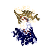
| ||||||||
|---|---|---|---|---|---|---|---|---|---|
| 1 | 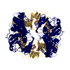
| ||||||||
| Unit cell |
| ||||||||
| Noncrystallographic symmetry (NCS) | NCS oper: (Code: given Matrix: (-1, 0.00212, -0.00057), Vector : Details | THE NON-CRYSTALLOGRAPHIC DIAD RELATING THE TWO SUBUNITS IS GIVEN ON THE MTRIX RECORDS BELOW. APPLYING THIS TRANSFORMATION TO THE RED SUBUNIT WILL YIELD APPROXIMATE COORDINATES FOR THE GREEN SUBUNIT. THIS DIAD CORRESPONDS CLOSELY TO THE CRYSTAL C-AXIS. THE TRANSFORMATION WAS DERIVED USING THE SYMMETRY AVERAGING PROCEDURE CONTAINED IN THE HENDRICKSON AND KONNERT PROGRAM. THIS CORRESPONDS TO THE R-AXIS OF THE P, Q, R SCHEME USED IN STRUCTURAL STUDIES OF OTHER SPECIES OF THE ENZYME. | |
-Components
| #1: Protein | Mass: 35922.945 Da / Num. of mol.: 2 Source method: isolated from a genetically manipulated source Source: (gene. exp.) Homo sapiens (human)References: UniProt: P00354, UniProt: P04406*PLUS, glyceraldehyde-3-phosphate dehydrogenase (phosphorylating)#2: Chemical | ChemComp-SO4 / Sulfate  Mass: 96.063 Da / Num. of mol.: 4 / Source method: obtained synthetically / Formula: SO4 Mass: 96.063 Da / Num. of mol.: 4 / Source method: obtained synthetically / Formula: SO4#3: Chemical | Nicotinamide adenine dinucleotide  Mass: 663.425 Da / Num. of mol.: 2 / Source method: obtained synthetically / Formula: C21H27N7O14P2 / Comment: NAD*YM Mass: 663.425 Da / Num. of mol.: 2 / Source method: obtained synthetically / Formula: C21H27N7O14P2 / Comment: NAD*YMCompound details | TURNS ARE SPECIFIED WHERE CA(N) TO CA(N+3) IS LESS THAN 5.7 ANGSTROMS AND O(N) TO N(N+3) IS LESS ...TURNS ARE SPECIFIED WHERE CA(N) TO CA(N+3) IS LESS THAN 5.7 ANGSTROMS AND O(N) TO N(N+3) IS LESS THAN 3.2 ANGSTROMS. MORE EXACT TURN DEFINITION | |
|---|
-Experimental details
-Experiment
| Experiment | Method: X-RAY DIFFRACTION |
|---|
- Sample preparation
| Crystal | Density Matthews: 3.35 Å3/Da / Density % sol: 63.32 % | |||||||||||||||||||||||||
|---|---|---|---|---|---|---|---|---|---|---|---|---|---|---|---|---|---|---|---|---|---|---|---|---|---|---|
| Crystal grow | *PLUS pH: 5.5 / Method: microdialysis | |||||||||||||||||||||||||
| Components of the solutions | *PLUS
|
- Processing
| Refinement | Rfactor Rwork: 0.33 / Highest resolution: 3.5 Å Details: THE RESOLUTION COULD BE EXTENDED TO ABOUT 1.5 ANGSTROMS USING SYNCHROTRON RADIATION. | ||||||||||||
|---|---|---|---|---|---|---|---|---|---|---|---|---|---|
| Refinement step | Cycle: LAST / Highest resolution: 3.5 Å
| ||||||||||||
| Refinement | *PLUS Highest resolution: 3.5 Å / Rfactor obs: 0.33 | ||||||||||||
| Solvent computation | *PLUS | ||||||||||||
| Displacement parameters | *PLUS |
