 Movie
Movie Controller

[English] 日本語
 Yorodumi
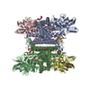
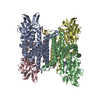
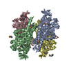
- PDB-3gnc: Crystal structure of Glutaryl-COA dehydrogenase from Burkholderia... -
+ Open data

- Basic information
| Entry | Database: PDB / ID: 3gnc | ||||||
|---|---|---|---|---|---|---|---|
| Title | Crystal structure of Glutaryl-COA dehydrogenase from Burkholderia Pseudomallei with fragment 6421 | ||||||
 Components Components | Glutaryl-CoA dehydrogenase | ||||||
 Keywords Keywords | OXIDOREDUCTASE / Structural Genomics / Seattle Structural Genomics Center for Infectious Disease / SSGCID | ||||||
| Function / homology |  Function and homology information Function and homology informationglutaryl-CoA dehydrogenase (ETF) / glutaryl-CoA dehydrogenase activity / flavin adenine dinucleotide bindingSimilarity search - Function | ||||||
| Biological species |  Burkholderia pseudomallei (bacteria) Burkholderia pseudomallei (bacteria) | ||||||
| Method | X-RAY DIFFRACTION / SYNCHROTRON / MOLECULAR REPLACEMENT / Resolution: 2.15 Å | ||||||
 Authors Authors | Seattle Structural Genomics Center for Infectious Disease (SSGCID) | ||||||
 Citation Citation | Journal: Acta Crystallogr.,Sect.F / Year: 2011 Title: Probing conformational states of glutaryl-CoA dehydrogenase by fragment screening. Authors: Begley, D.W. / Davies, D.R. / Hartley, R.C. / Hewitt, S.N. / Rychel, A.L. / Myler, P.J. / Van Voorhis, W.C. / Staker, B.L. / Stewart, L.J. | ||||||
| History |
|
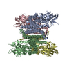
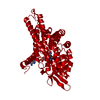
- Structure visualization
| Structure viewer | Molecule: MolmilJmol/JSmol |
|---|
- Downloads & links
-Download
| PDBx/mmCIF format | 3gnc.cif.gz | 294.9 KB | Display | PDBx/mmCIF format |
|---|---|---|---|---|
| PDB format | pdb3gnc.ent.gz | 239.2 KB | Display | PDB format |
| PDBx/mmJSON format | 3gnc.json.gz | Tree view | PDBx/mmJSON format | |
| Others |  Other downloads Other downloads |
-Validation report
| Arichive directory | https://data.pdbj.org/pub/pdb/validation_reports/gn/3gncftp://data.pdbj.org/pub/pdb/validation_reports/gn/3gnc | HTTPS FTP |
|---|
-Related structure data
| Related structure data |  3d6bSC  3eomC 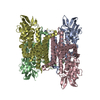 3eonC 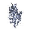 3gqtC S: Starting model for refinement C: citing same article ( |
|---|---|
| Similar structure data | |
| Other databases |

-Links
 PDBj
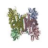
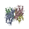
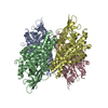
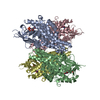
PDBj- Assembly
| Deposited unit | 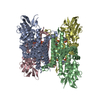
| ||||||||
|---|---|---|---|---|---|---|---|---|---|
| 1 |
| ||||||||
| Unit cell |
|
-Components
| #1: Protein | Mass: 43467.543 Da / Num. of mol.: 4 Source method: isolated from a genetically manipulated source Source: (gene. exp.) Burkholderia pseudomallei (bacteria) / Strain: 1710b / Gene: BURPS1710b_3237 / Plasmid: AVA0421 / Production host: Escherichia coli (E. coli) / Strain (production host): BL21(DE3) / References: UniProt: Q3JP94, glutaryl-CoA dehydrogenase#2: Chemical | ChemComp-SO4 / | Sulfate  Mass: 96.063 Da / Num. of mol.: 1 / Source method: obtained synthetically / Formula: SO4 Mass: 96.063 Da / Num. of mol.: 1 / Source method: obtained synthetically / Formula: SO4#3: Chemical | ChemComp-QQQ / 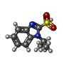  Mass: 240.279 Da / Num. of mol.: 4 / Source method: obtained synthetically / Formula: C10H12N2O3S Mass: 240.279 Da / Num. of mol.: 4 / Source method: obtained synthetically / Formula: C10H12N2O3S#4: Chemical | HEPES  Mass: 238.305 Da / Num. of mol.: 3 / Source method: obtained synthetically / Formula: C8H18N2O4S / Comment: pH buffer*YM Mass: 238.305 Da / Num. of mol.: 3 / Source method: obtained synthetically / Formula: C8H18N2O4S / Comment: pH buffer*YM#5: Water | ChemComp-HOH / | Water Mass: 18.015 Da / Num. of mol.: 210 / Source method: isolated from a natural source / Formula: H2O Mass: 18.015 Da / Num. of mol.: 210 / Source method: isolated from a natural source / Formula: H2O |
|---|
-Experimental details
-Experiment
| Experiment | Method: X-RAY DIFFRACTION / Number of used crystals: 1 |
|---|
- Sample preparation
| Crystal | Density Matthews: 2.16 Å3/Da / Density % sol: 43.1 % |
|---|---|
| Crystal grow | Temperature: 298 K / Method: vapor diffusion, sitting drop / pH: 7.5 Details: 20% PEG 3000, 0.1M HEPES, 0.2M NaCl, pH 7.5, VAPOR DIFFUSION, SITTING DROP, temperature 298K |
-Data collection
| Diffraction | Mean temperature: 100 K |
|---|---|
| Diffraction source | Source: SYNCHROTRON / Site: ALS  / Beamline: 5.0.3 / Wavelength: 0.97 / Beamline: 5.0.3 / Wavelength: 0.97 |
| Detector | Type: ADSC QUANTUM 315 / Detector: CCD / Date: Mar 1, 2009 |
| Radiation | Protocol: SINGLE WAVELENGTH / Monochromatic (M) / Laue (L): M / Scattering type: x-ray |
| Radiation wavelength | Wavelength: 0.97 Å / Relative weight: 1 |
| Reflection | Resolution: 2.15→50 Å / Num. obs: 81993 / % possible obs: 99.6 % / Redundancy: 7 % / Rmerge(I) obs: 0.072 / Net I/σ(I): 10.8 |
| Reflection shell | Resolution: 2.15→2.19 Å / Redundancy: 6.8 % / Rmerge(I) obs: 0.593 / % possible all: 98.5 |
- Processing
| Software |
| ||||||||||||||||||||||||||||||||||||||||||||||||||||||||||||||||||||||||||||||||||||||||||||||||||||||||||||||||||||||||||||||||||||||||||||||||||||||||||||||||||||||||||
|---|---|---|---|---|---|---|---|---|---|---|---|---|---|---|---|---|---|---|---|---|---|---|---|---|---|---|---|---|---|---|---|---|---|---|---|---|---|---|---|---|---|---|---|---|---|---|---|---|---|---|---|---|---|---|---|---|---|---|---|---|---|---|---|---|---|---|---|---|---|---|---|---|---|---|---|---|---|---|---|---|---|---|---|---|---|---|---|---|---|---|---|---|---|---|---|---|---|---|---|---|---|---|---|---|---|---|---|---|---|---|---|---|---|---|---|---|---|---|---|---|---|---|---|---|---|---|---|---|---|---|---|---|---|---|---|---|---|---|---|---|---|---|---|---|---|---|---|---|---|---|---|---|---|---|---|---|---|---|---|---|---|---|---|---|---|---|---|---|---|---|---|
| Refinement | Method to determine structure: MOLECULAR REPLACEMENT Starting model: pdb entry 3D6B Resolution: 2.15→46.68 Å / Cor.coef. Fo:Fc: 0.955 / Cor.coef. Fo:Fc free: 0.93 / SU B: 6.495 / SU ML: 0.168 / Cross valid method: THROUGHOUT / σ(F): 0 / ESU R: 0.283 / ESU R Free: 0.218 / Stereochemistry target values: MAXIMUM LIKELIHOOD / Details: HYDROGENS HAVE BEEN ADDED IN THE
| ||||||||||||||||||||||||||||||||||||||||||||||||||||||||||||||||||||||||||||||||||||||||||||||||||||||||||||||||||||||||||||||||||||||||||||||||||||||||||||||||||||||||||
| Solvent computation | Ion probe radii: 0.8 Å / Shrinkage radii: 0.8 Å / VDW probe radii: 1.2 Å / Solvent model: MASK | ||||||||||||||||||||||||||||||||||||||||||||||||||||||||||||||||||||||||||||||||||||||||||||||||||||||||||||||||||||||||||||||||||||||||||||||||||||||||||||||||||||||||||
| Displacement parameters | Biso mean: 37.44 Å2
| ||||||||||||||||||||||||||||||||||||||||||||||||||||||||||||||||||||||||||||||||||||||||||||||||||||||||||||||||||||||||||||||||||||||||||||||||||||||||||||||||||||||||||
| Refinement step | Cycle: LAST / Resolution: 2.15→46.68 Å
| ||||||||||||||||||||||||||||||||||||||||||||||||||||||||||||||||||||||||||||||||||||||||||||||||||||||||||||||||||||||||||||||||||||||||||||||||||||||||||||||||||||||||||
| Refine LS restraints |
| ||||||||||||||||||||||||||||||||||||||||||||||||||||||||||||||||||||||||||||||||||||||||||||||||||||||||||||||||||||||||||||||||||||||||||||||||||||||||||||||||||||||||||
| LS refinement shell | Resolution: 2.15→2.21 Å / Total num. of bins used: 20
|
