 Movie
Movie Controller

[English] 日本語
 Yorodumi
- PDB-3gmt: Crystal structure of adenylate kinase from burkholderia pseudomallei -
+ Open data

- Basic information
| Entry | Database: PDB / ID: 3gmt | ||||||
|---|---|---|---|---|---|---|---|
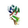
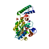
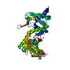
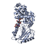
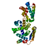
| Title | Crystal structure of adenylate kinase from burkholderia pseudomallei | ||||||
 Components Components | Adenylate kinase | ||||||
 Keywords Keywords | TRANSFERASE / SSGCID / ADENYLATE KINASE / BURKHOLDERIA PSEUDOMALLEI / ATP-binding / Kinase / Nucleotide biosynthesis / Nucleotide-binding / Structural Genomics / Seattle Structural Genomics Center for Infectious Disease | ||||||
| Function / homology |  Function and homology informationadenylate kinase / adenylate kinase activity / AMP salvage / ATP binding / cytoplasm Function and homology informationadenylate kinase / adenylate kinase activity / AMP salvage / ATP binding / cytoplasmSimilarity search - Function | ||||||
| Biological species |  Burkholderia pseudomallei 1710b (bacteria) Burkholderia pseudomallei 1710b (bacteria) | ||||||
| Method | X-RAY DIFFRACTION / SYNCHROTRON / SAD / Resolution: 2.1 Å | ||||||
 Authors Authors | Abendroth, J. / Staker, B.L. / Robinson, H. / Buchko, G.W. / Hewitt, S.N. / Napuli, A.J. / Van Voorhis, W. / Stacy, R. / Myler, P.J. / Stewart, L. / Seattle Structural Genomics Center for Infectious Disease (SSGCID) | ||||||
 Citation Citation | Journal: Biochem.Biophys.Res.Commun. / Year: 2010 Title: Structural characterization of Burkholderia pseudomallei adenylate kinase (Adk): profound asymmetry in the crystal structure of the 'open' state. Authors: Buchko, G.W. / Robinson, H. / Abendroth, J. / Staker, B.L. / Myler, P.J. #1: Journal: Plos One / Year: 2013Title: Combining functional and structural genomics to sample the essential Burkholderia structome. Authors: Baugh, L. / Gallagher, L.A. / Patrapuvich, R. / Clifton, M.C. / Gardberg, A.S. / Edwards, T.E. / Armour, B. / Begley, D.W. / Dieterich, S.H. / Dranow, D.M. / Abendroth, J. / Fairman, J.W. / ...Authors: Baugh, L. / Gallagher, L.A. / Patrapuvich, R. / Clifton, M.C. / Gardberg, A.S. / Edwards, T.E. / Armour, B. / Begley, D.W. / Dieterich, S.H. / Dranow, D.M. / Abendroth, J. / Fairman, J.W. / Fox, D. / Staker, B.L. / Phan, I. / Gillespie, A. / Choi, R. / Nakazawa-Hewitt, S. / Nguyen, M.T. / Napuli, A. / Barrett, L. / Buchko, G.W. / Stacy, R. / Myler, P.J. / Stewart, L.J. / Manoil, C. / Van Voorhis, W.C. | ||||||
| History |
|
- Structure visualization
| Structure viewer | Molecule: MolmilJmol/JSmol |
|---|
- Downloads & links
-Download
| PDBx/mmCIF format | 3gmt.cif.gz | 91.5 KB | Display | PDBx/mmCIF format |
|---|---|---|---|---|
| PDB format | pdb3gmt.ent.gz | 73.9 KB | Display | PDB format |
| PDBx/mmJSON format | 3gmt.json.gz | Tree view | PDBx/mmJSON format | |
| Others |  Other downloads Other downloads |
-Validation report
| Arichive directory | https://data.pdbj.org/pub/pdb/validation_reports/gm/3gmtftp://data.pdbj.org/pub/pdb/validation_reports/gm/3gmt | HTTPS FTP |
|---|
-Related structure data
| Similar structure data | |
|---|---|
| Other databases |


-Links
 PDBj
PDBj- Assembly
| Deposited unit | 
| ||||||||
|---|---|---|---|---|---|---|---|---|---|
| 1 | 
| ||||||||
| 2 | 
| ||||||||
| Unit cell |
|
-Components
| #1: Protein | / AK / ATP-AMP transphosphorylase Mass: 25771.525 Da / Num. of mol.: 2 Source method: isolated from a genetically manipulated source Source: (gene. exp.) Burkholderia pseudomallei 1710b (bacteria)Strain: 1719B / Gene: adk, BURPS1710b_1080 / Plasmid: AVA0421 / Production host: Escherichia coli (E. coli) / Strain (production host): BL21(DE3) / References: UniProt: Q3JVB1, adenylate kinase#2: Chemical | ChemComp-SO4 / Sulfate  Mass: 96.063 Da / Num. of mol.: 4 / Source method: obtained synthetically / Formula: SO4 Mass: 96.063 Da / Num. of mol.: 4 / Source method: obtained synthetically / Formula: SO4#3: Water | ChemComp-HOH / | Water Mass: 18.015 Da / Num. of mol.: 216 / Source method: isolated from a natural source / Formula: H2O Mass: 18.015 Da / Num. of mol.: 216 / Source method: isolated from a natural source / Formula: H2O |
|---|
-Experimental details
-Experiment
| Experiment | Method: X-RAY DIFFRACTION / Number of used crystals: 1 |
|---|
- Sample preparation
| Crystal | Density Matthews: 2.27 Å3/Da / Density % sol: 46 % |
|---|---|
| Crystal grow | Temperature: 290 K / Method: vapor diffusion, sitting drop / pH: 8 Details: HAMPTON SCREEN, B5: 30% PEG 4000, 200MM LI2SO4, 100MM TRIS PH 8,, VAPOR DIFFUSION, SITTING DROP, temperature 290K |
-Data collection
| Diffraction | Mean temperature: 100 K |
|---|---|
| Diffraction source | Source: SYNCHROTRON / Site: NSLS  / Beamline: X8C / Wavelength: 0.9791 / Wavelength: 0.9791 Å / Beamline: X8C / Wavelength: 0.9791 / Wavelength: 0.9791 Å |
| Detector | Type: ADSC QUANTUM Q315r / Detector: CCD / Date: Jan 31, 2009 |
| Radiation | Protocol: SINGLE WAVELENGTH / Monochromatic (M) / Laue (L): M / Scattering type: x-ray |
| Radiation wavelength | Wavelength: 0.9791 Å / Relative weight: 1 |
| Reflection | Resolution: 2.1→20 Å / Num. all: 27839 / Num. obs: 27703 / % possible obs: 99.5 % / Observed criterion σ(F): 0 / Observed criterion σ(I): -3 / Redundancy: 7.4 % / Biso Wilson estimate: 42.51 Å2 / Rmerge(I) obs: 0.072 / Net I/σ(I): 17.02 |
| Reflection shell | Resolution: 2.1→2.15 Å / Redundancy: 7.6 % / Rmerge(I) obs: 0.618 / Mean I/σ(I) obs: 3.1 / Num. unique all: 2051 / Rsym value: 0.618 / % possible all: 99.8 |
- Processing
| Software |
| ||||||||||||||||||||||||||||||||||||||||||||||||||||||||||||||||||||||||||||||||||||||||||||||||||||||||||||||||||||||||||||||||||||||||||||||||||||||||||||||||||||||||||
|---|---|---|---|---|---|---|---|---|---|---|---|---|---|---|---|---|---|---|---|---|---|---|---|---|---|---|---|---|---|---|---|---|---|---|---|---|---|---|---|---|---|---|---|---|---|---|---|---|---|---|---|---|---|---|---|---|---|---|---|---|---|---|---|---|---|---|---|---|---|---|---|---|---|---|---|---|---|---|---|---|---|---|---|---|---|---|---|---|---|---|---|---|---|---|---|---|---|---|---|---|---|---|---|---|---|---|---|---|---|---|---|---|---|---|---|---|---|---|---|---|---|---|---|---|---|---|---|---|---|---|---|---|---|---|---|---|---|---|---|---|---|---|---|---|---|---|---|---|---|---|---|---|---|---|---|---|---|---|---|---|---|---|---|---|---|---|---|---|---|---|---|
| Refinement | Method to determine structure: SAD / Resolution: 2.1→20 Å / Cor.coef. Fo:Fc: 0.938 / Cor.coef. Fo:Fc free: 0.901 / SU B: 6.724 / SU ML: 0.179 / Isotropic thermal model: isotropic / Cross valid method: THROUGHOUT / σ(F): 0 / σ(I): 0 / ESU R: 0.253 / ESU R Free: 0.224 / Stereochemistry target values: MAXIMUM LIKELIHOOD / Details: HYDROGENS HAVE BEEN ADDED IN THE
| ||||||||||||||||||||||||||||||||||||||||||||||||||||||||||||||||||||||||||||||||||||||||||||||||||||||||||||||||||||||||||||||||||||||||||||||||||||||||||||||||||||||||||
| Solvent computation | Ion probe radii: 0.8 Å / Shrinkage radii: 0.8 Å / VDW probe radii: 1.4 Å / Solvent model: MASK | ||||||||||||||||||||||||||||||||||||||||||||||||||||||||||||||||||||||||||||||||||||||||||||||||||||||||||||||||||||||||||||||||||||||||||||||||||||||||||||||||||||||||||
| Displacement parameters | Biso mean: 29.82 Å2
| ||||||||||||||||||||||||||||||||||||||||||||||||||||||||||||||||||||||||||||||||||||||||||||||||||||||||||||||||||||||||||||||||||||||||||||||||||||||||||||||||||||||||||
| Refinement step | Cycle: LAST / Resolution: 2.1→20 Å
| ||||||||||||||||||||||||||||||||||||||||||||||||||||||||||||||||||||||||||||||||||||||||||||||||||||||||||||||||||||||||||||||||||||||||||||||||||||||||||||||||||||||||||
| Refine LS restraints |
| ||||||||||||||||||||||||||||||||||||||||||||||||||||||||||||||||||||||||||||||||||||||||||||||||||||||||||||||||||||||||||||||||||||||||||||||||||||||||||||||||||||||||||
| LS refinement shell | Resolution: 2.1→2.15 Å / Total num. of bins used: 20
|
