 Movie
Movie Controller


+ Open data

- Basic information
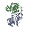
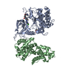
| Entry | Database: PDB / ID: 3glu | ||||||
|---|---|---|---|---|---|---|---|
| Title | Crystal Structure of Human SIRT3 with AceCS2 peptide | ||||||
 Components Components |
| ||||||
 Keywords Keywords | HYDROLASE/HYDROLASE REGULATOR / NAD dependent deacetylase /  sirtuin / product peptide complex / Hydrolase / Metal-binding / Mitochondrion / NAD / Polymorphism / Transit peptide / Zinc / Alternative splicing / Ligase / HYDROLASE-HYDROLASE REGULATOR COMPLEX sirtuin / product peptide complex / Hydrolase / Metal-binding / Mitochondrion / NAD / Polymorphism / Transit peptide / Zinc / Alternative splicing / Ligase / HYDROLASE-HYDROLASE REGULATOR COMPLEX | ||||||
| Function / homology |  Function and homology information Function and homology informationpropionate biosynthetic process / acetate biosynthetic process / propionate-CoA ligase / propionate-CoA ligase activity / positive regulation of catalase activity / positive regulation of ceramide biosynthetic process / acetate-CoA ligase / acetate-CoA ligase activity / acetyl-CoA biosynthetic process from acetate / Ethanol oxidation ...propionate biosynthetic process / acetate biosynthetic process / propionate-CoA ligase / propionate-CoA ligase activity / positive regulation of catalase activity / positive regulation of ceramide biosynthetic process / acetate-CoA ligase / acetate-CoA ligase activity / acetyl-CoA biosynthetic process from acetate / Ethanol oxidation / peptidyl-lysine deacetylation / acetyl-CoA biosynthetic process / positive regulation of superoxide dismutase activity / : / NAD-dependent protein lysine deacetylase activity / protein acetyllysine N-acetyltransferase / NAD-dependent histone deacetylase activity / protein deacetylation / Regulation of FOXO transcriptional activity by acetylation / AMP binding / NAD+ binding / negative regulation of reactive oxygen species metabolic process / FOXO-mediated transcription of oxidative stress, metabolic and neuronal genes / aerobic respiration / Transcriptional activation of mitochondrial biogenesis / positive regulation of insulin secretion / negative regulation of ERK1 and ERK2 cascade / transferase activity / sequence-specific DNA binding / mitochondrial matrix / enzyme binding / protein-containing complex / mitochondrion / zinc ion binding / nucleoplasm / ATP binding / nucleusSimilarity search - Function | ||||||
| Biological species |  Homo sapiens (human) Homo sapiens (human) | ||||||
| Method | X-RAY DIFFRACTION / SYNCHROTRON / MOLECULAR REPLACEMENT / Resolution: 2.5 Å | ||||||
 Authors Authors | Jin, L. / Wei, W. / Jiang, Y. / Peng, H. / Cai, J. / Mao, C. / Dai, H. / Bemis, J.E. / Jirousek, M.R. / Milne, J.C. ...Jin, L. / Wei, W. / Jiang, Y. / Peng, H. / Cai, J. / Mao, C. / Dai, H. / Bemis, J.E. / Jirousek, M.R. / Milne, J.C. / Westphal, C.H. / Perni, R.B. | ||||||
 Citation Citation | Journal: J.Biol.Chem. / Year: 2009 Title: Crystal Structures of Human SIRT3 Displaying Substrate-induced Conformational Changes. Authors: Jin, L. / Wei, W. / Jiang, Y. / Peng, H. / Cai, J. / Mao, C. / Dai, H. / Choy, W. / Bemis, J.E. / Jirousek, M.R. / Milne, J.C. / Westphal, C.H. / Perni, R.B. | ||||||
| History |
|
- Structure visualization
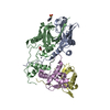
| Structure viewer | Molecule: MolmilJmol/JSmol |
|---|
- Downloads & links
-Download
| PDBx/mmCIF format | 3glu.cif.gz | 70.1 KB | Display | PDBx/mmCIF format |
|---|---|---|---|---|
| PDB format | pdb3glu.ent.gz | 50.3 KB | Display | PDB format |
| PDBx/mmJSON format | 3glu.json.gz | Tree view | PDBx/mmJSON format | |
| Others |  Other downloads Other downloads |
-Validation report
| Arichive directory | https://data.pdbj.org/pub/pdb/validation_reports/gl/3gluftp://data.pdbj.org/pub/pdb/validation_reports/gl/3glu | HTTPS FTP |
|---|
-Related structure data
| Related structure data | 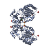 3glrSC  3glsC 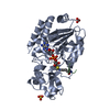 3gltC S: Starting model for refinement C: citing same article ( |
|---|---|
| Similar structure data |


-Links
 PDBj
PDBj


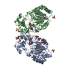
- Assembly
| Deposited unit | 
| ||||||||
|---|---|---|---|---|---|---|---|---|---|
| 1 | 
| ||||||||
| Unit cell |
|
-Components
| #1: Protein | Mass: 31612.348 Da / Num. of mol.: 1 / Fragment: Human SIRT3, residues 118-399 Source method: isolated from a genetically manipulated source Source: (gene. exp.) Homo sapiens (human) / Gene: SIRT3, SIR2L3 / Plasmid: modified pET21b / Production host:  Escherichia coli (E. coli) / Strain (production host): BL21-GOLD(DE3) Escherichia coli (E. coli) / Strain (production host): BL21-GOLD(DE3)References: UniProt: Q9NTG7, Hydrolases; Acting on carbon-nitrogen bonds, other than peptide bonds; In linear amides | ||||
|---|---|---|---|---|---|
| #2: Protein/peptide | Mass: 1477.846 Da / Num. of mol.: 1 / Fragment: Human Acyl-CoA, residues 638-649 / Source method: obtained synthetically / Source: (synth.) Homo sapiens (human) / References: UniProt: Q9NUB1, acetate-CoA ligase | ||||
| #3: Chemical | Sulfate  Mass: 96.063 Da / Num. of mol.: 2 / Source method: obtained synthetically / Formula: SO4 Mass: 96.063 Da / Num. of mol.: 2 / Source method: obtained synthetically / Formula: SO4#4: Chemical | ChemComp-ZN / |   Mass: 65.409 Da / Num. of mol.: 1 / Source method: obtained synthetically / Formula: Zn Mass: 65.409 Da / Num. of mol.: 1 / Source method: obtained synthetically / Formula: Zn#5: Water | ChemComp-HOH / | Water Mass: 18.015 Da / Num. of mol.: 68 / Source method: isolated from a natural source / Formula: H2O Mass: 18.015 Da / Num. of mol.: 68 / Source method: isolated from a natural source / Formula: H2O |
-Experimental details
-Experiment
| Experiment | Method: X-RAY DIFFRACTION / Number of used crystals: 1 |
|---|
- Sample preparation
| Crystal | Density Matthews: 2.98 Å3/Da / Density % sol: 58.71 % |
|---|---|
| Crystal grow | Temperature: 291 K / Method: vapor diffusion, hanging drop / pH: 5.5 Details: 0.2 M lithium sulfate monohydrate, 17% w/v PEG 12000 and 0.1 M Bis-Tris, pH 5.5, VAPOR DIFFUSION, HANGING DROP, temperature 291K |
-Data collection
| Diffraction | Mean temperature: 100 K |
|---|---|
| Diffraction source | Source: SYNCHROTRON / Site: APS  / Beamline: 24-ID-E / Wavelength: 0.97918 Å / Beamline: 24-ID-E / Wavelength: 0.97918 Å |
| Detector | Type: ADSC QUANTUM 315 / Detector: CCD / Date: Oct 4, 2008 |
| Radiation | Monochromator: Cryogenically-cooled single crystal Si(111) side bounce monochromator Protocol: SINGLE WAVELENGTH / Monochromatic (M) / Laue (L): M / Scattering type: x-ray |
| Radiation wavelength | Wavelength: 0.97918 Å / Relative weight: 1 |
| Reflection | Resolution: 2.5→65 Å / Num. obs: 13322 / % possible obs: 95.7 % / Observed criterion σ(I): 2 / Redundancy: 4.1 % / Rmerge(I) obs: 0.087 / Net I/σ(I): 10.3 |
| Reflection shell | Resolution: 2.5→2.64 Å / Redundancy: 4.2 % / Rmerge(I) obs: 0.446 / Mean I/σ(I) obs: 3.3 / Num. unique all: 1942 / % possible all: 96.9 |
- Processing
| Software |
| ||||||||||||||||||||||||||||||||||||||||||||||||||||||||||||||||||||||||||||||||||||||||||||||||||||||||||||||||||||||||||||||||||||||||||||||||||||||||||||||||||||||||||
|---|---|---|---|---|---|---|---|---|---|---|---|---|---|---|---|---|---|---|---|---|---|---|---|---|---|---|---|---|---|---|---|---|---|---|---|---|---|---|---|---|---|---|---|---|---|---|---|---|---|---|---|---|---|---|---|---|---|---|---|---|---|---|---|---|---|---|---|---|---|---|---|---|---|---|---|---|---|---|---|---|---|---|---|---|---|---|---|---|---|---|---|---|---|---|---|---|---|---|---|---|---|---|---|---|---|---|---|---|---|---|---|---|---|---|---|---|---|---|---|---|---|---|---|---|---|---|---|---|---|---|---|---|---|---|---|---|---|---|---|---|---|---|---|---|---|---|---|---|---|---|---|---|---|---|---|---|---|---|---|---|---|---|---|---|---|---|---|---|---|---|---|
| Refinement | Method to determine structure: MOLECULAR REPLACEMENT Starting model: 3GLR Resolution: 2.5→65 Å / Cor.coef. Fo:Fc: 0.935 / Cor.coef. Fo:Fc free: 0.919 / Isotropic thermal model: Isotropic / Cross valid method: THROUGHOUT / ESU R: 0.419 / ESU R Free: 0.271 / Stereochemistry target values: MAXIMUM LIKELIHOOD
| ||||||||||||||||||||||||||||||||||||||||||||||||||||||||||||||||||||||||||||||||||||||||||||||||||||||||||||||||||||||||||||||||||||||||||||||||||||||||||||||||||||||||||
| Solvent computation | Ion probe radii: 0.8 Å / Shrinkage radii: 0.8 Å / VDW probe radii: 1.4 Å / Solvent model: MASK | ||||||||||||||||||||||||||||||||||||||||||||||||||||||||||||||||||||||||||||||||||||||||||||||||||||||||||||||||||||||||||||||||||||||||||||||||||||||||||||||||||||||||||
| Displacement parameters | Biso mean: 39.224 Å2
| ||||||||||||||||||||||||||||||||||||||||||||||||||||||||||||||||||||||||||||||||||||||||||||||||||||||||||||||||||||||||||||||||||||||||||||||||||||||||||||||||||||||||||
| Refinement step | Cycle: LAST / Resolution: 2.5→65 Å
| ||||||||||||||||||||||||||||||||||||||||||||||||||||||||||||||||||||||||||||||||||||||||||||||||||||||||||||||||||||||||||||||||||||||||||||||||||||||||||||||||||||||||||
| Refine LS restraints |
| ||||||||||||||||||||||||||||||||||||||||||||||||||||||||||||||||||||||||||||||||||||||||||||||||||||||||||||||||||||||||||||||||||||||||||||||||||||||||||||||||||||||||||
| LS refinement shell | Resolution: 2.5→2.565 Å / Total num. of bins used: 20
|
