 Movie
Movie Controller


+ Open data

- Basic information
| Entry | Database: PDB / ID: 3gj0 | ||||||
|---|---|---|---|---|---|---|---|
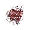
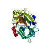
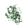
| Title | Crystal structure of human RanGDP | ||||||
 Components Components | GTP-binding nuclear protein Ran | ||||||
 Keywords Keywords |  TRANSPORT PROTEIN / G Protein / GDP / Acetylation / Cytoplasm / GTP-binding / Host-virus interaction / Nucleotide-binding / Nucleus / Phosphoprotein / Polymorphism / Protein transport / Transport TRANSPORT PROTEIN / G Protein / GDP / Acetylation / Cytoplasm / GTP-binding / Host-virus interaction / Nucleotide-binding / Nucleus / Phosphoprotein / Polymorphism / Protein transport / Transport | ||||||
| Function / homology |  Function and homology information Function and homology informationRNA nuclear export complex / pre-miRNA export from nucleus / snRNA import into nucleus / cellular response to mineralocorticoid stimulus / manchette / Regulation of cholesterol biosynthesis by SREBP (SREBF) / importin-alpha family protein binding / protein localization to nucleolus / Rev-mediated nuclear export of HIV RNA / Nuclear import of Rev protein ...RNA nuclear export complex / pre-miRNA export from nucleus / snRNA import into nucleus / cellular response to mineralocorticoid stimulus / manchette / Regulation of cholesterol biosynthesis by SREBP (SREBF) / importin-alpha family protein binding / protein localization to nucleolus / Rev-mediated nuclear export of HIV RNA / Nuclear import of Rev protein / GTP metabolic process / NEP/NS2 Interacts with the Cellular Export Machinery / tRNA processing in the nucleus / Postmitotic nuclear pore complex (NPC) reformation / MicroRNA (miRNA) biogenesis / dynein intermediate chain binding / DNA metabolic process / mitotic sister chromatid segregation / spermatid development / ribosomal large subunit export from nucleus / sperm flagellum / ribosomal small subunit export from nucleus / ribosomal subunit export from nucleus / nuclear pore / centriole / protein export from nucleus / viral process / mitotic spindle organization / G protein activity / male germ cell nucleus / hippocampus development / Transcriptional regulation by small RNAs / Hydrolases; Acting on acid anhydrides; Acting on GTP to facilitate cellular and subcellular movement / recycling endosome / positive regulation of protein import into nucleus / protein import into nucleus / GDP binding / melanosome / positive regulation of protein binding / nuclear envelope / mitotic cell cycle / midbody / actin cytoskeleton organization / cadherin binding / protein heterodimerization activity / protein domain specific binding / cell division / GTPase activity / chromatin binding / chromatin / nucleolus / GTP binding / magnesium ion binding / protein-containing complex / RNA binding / extracellular exosome / nucleoplasm / membrane / nucleus / cytosol / cytoplasmSimilarity search - Function | ||||||
| Biological species |  Homo sapiens (human) Homo sapiens (human) | ||||||
| Method | X-RAY DIFFRACTION / SYNCHROTRON / MOLECULAR REPLACEMENT / Resolution: 1.48 Å | ||||||
 Authors Authors | Partridge, J.R. / Schwartz, T.U. | ||||||
 Citation Citation | Journal: J.Mol.Biol. / Year: 2009 Title: Crystallographic and Biochemical Analysis of the Ran-binding Zinc Finger Domain. Authors: Partridge, J.R. / Schwartz, T.U. | ||||||
| History |
|
- Structure visualization
| Structure viewer | Molecule: MolmilJmol/JSmol |
|---|
- Downloads & links
-Download
| PDBx/mmCIF format | 3gj0.cif.gz | 197.5 KB | Display | PDBx/mmCIF format |
|---|---|---|---|---|
| PDB format | pdb3gj0.ent.gz | 156 KB | Display | PDB format |
| PDBx/mmJSON format | 3gj0.json.gz | Tree view | PDBx/mmJSON format | |
| Others |  Other downloads Other downloads |
-Validation report
| Arichive directory | https://data.pdbj.org/pub/pdb/validation_reports/gj/3gj0ftp://data.pdbj.org/pub/pdb/validation_reports/gj/3gj0 | HTTPS FTP |
|---|
-Related structure data
| Related structure data |  3gj3C  3gj4C  3gj5C  3gj6C  3gj7C  3gj8C  1byuS S: Starting model for refinement C: citing same article ( |
|---|---|
| Similar structure data |


-Links
 PDBj
PDBj


- Assembly
| Deposited unit | 
| ||||||||
|---|---|---|---|---|---|---|---|---|---|
| 1 | 
| ||||||||
| 2 | 
| ||||||||
| Unit cell |
| ||||||||
| Components on special symmetry positions |
|
-Components
| #1: Protein | Mass: 24906.576 Da / Num. of mol.: 2 Source method: isolated from a genetically manipulated source Source: (gene. exp.) Homo sapiens (human) / Gene: ARA24, OK/SW-cl.81, RAN / Plasmid: pET28a / Production host:  Escherichia coli (E. coli) / Strain (production host): BL21(DE3)-RIL / References: UniProt: P62826 Escherichia coli (E. coli) / Strain (production host): BL21(DE3)-RIL / References: UniProt: P62826#2: Chemical |   Mass: 24.305 Da / Num. of mol.: 2 / Source method: obtained synthetically / Formula: Mg Mass: 24.305 Da / Num. of mol.: 2 / Source method: obtained synthetically / Formula: Mg#3: Chemical | Guanosine diphosphate  Type: RNA linking / Mass: 443.201 Da / Num. of mol.: 2 / Source method: obtained synthetically / Formula: C10H15N5O11P2 / Comment: GDP, energy-carrying molecule*YM Type: RNA linking / Mass: 443.201 Da / Num. of mol.: 2 / Source method: obtained synthetically / Formula: C10H15N5O11P2 / Comment: GDP, energy-carrying molecule*YM#4: Water | ChemComp-HOH / | Water Mass: 18.015 Da / Num. of mol.: 478 / Source method: isolated from a natural source / Formula: H2O Mass: 18.015 Da / Num. of mol.: 478 / Source method: isolated from a natural source / Formula: H2O |
|---|
-Experimental details
-Experiment
| Experiment | Method: X-RAY DIFFRACTION / Number of used crystals: 1 |
|---|
- Sample preparation
| Crystal | Density Matthews: 2.18 Å3/Da / Density % sol: 43.54 % |
|---|---|
| Crystal grow | Temperature: 295 K / Method: vapor diffusion, hanging drop / pH: 6.5 Details: 0.1M Bis-Tris pH 6.5, 18-20% PEG 3350, VAPOR DIFFUSION, HANGING DROP, temperature 295K |
-Data collection
| Diffraction | Mean temperature: 100 K |
|---|---|
| Diffraction source | Source: SYNCHROTRON / Site: APS  / Beamline: 24-ID-C / Wavelength: 0.979 Å / Beamline: 24-ID-C / Wavelength: 0.979 Å |
| Detector | Type: ADSC QUANTUM 315 / Detector: CCD / Date: Aug 4, 2008 |
| Radiation | Protocol: SINGLE WAVELENGTH / Monochromatic (M) / Laue (L): M / Scattering type: x-ray |
| Radiation wavelength | Wavelength: 0.979 Å / Relative weight: 1 |
| Reflection | Resolution: 1.48→50 Å / Num. all: 70774 / Num. obs: 70774 / % possible obs: 95.5 % / Observed criterion σ(F): 0 / Observed criterion σ(I): 0 / Redundancy: 9.6 % / Biso Wilson estimate: 19.6 Å2 / Rsym value: 0.07 / Net I/σ(I): 32.6 |
| Reflection shell | Resolution: 1.48→1.51 Å / Redundancy: 7.5 % / Mean I/σ(I) obs: 1.6 / % possible all: 93 |
- Processing
| Software |
| ||||||||||||||||||||||||||||||||||||||||||||||||||||||||||||||||||||||||||||||||||||||||||||||||||||||||||||||||||||||||||||||||||||||||||||||||||||||||||||||||||||||||||||||||||||||
|---|---|---|---|---|---|---|---|---|---|---|---|---|---|---|---|---|---|---|---|---|---|---|---|---|---|---|---|---|---|---|---|---|---|---|---|---|---|---|---|---|---|---|---|---|---|---|---|---|---|---|---|---|---|---|---|---|---|---|---|---|---|---|---|---|---|---|---|---|---|---|---|---|---|---|---|---|---|---|---|---|---|---|---|---|---|---|---|---|---|---|---|---|---|---|---|---|---|---|---|---|---|---|---|---|---|---|---|---|---|---|---|---|---|---|---|---|---|---|---|---|---|---|---|---|---|---|---|---|---|---|---|---|---|---|---|---|---|---|---|---|---|---|---|---|---|---|---|---|---|---|---|---|---|---|---|---|---|---|---|---|---|---|---|---|---|---|---|---|---|---|---|---|---|---|---|---|---|---|---|---|---|---|---|
| Refinement | Method to determine structure: MOLECULAR REPLACEMENT Starting model: PDB entry 1BYU Resolution: 1.48→43.217 Å / SU ML: 0.48 / σ(F): 1.33 / Phase error: 19.38 / Stereochemistry target values: MAXIMUM LIKELIHOOD
| ||||||||||||||||||||||||||||||||||||||||||||||||||||||||||||||||||||||||||||||||||||||||||||||||||||||||||||||||||||||||||||||||||||||||||||||||||||||||||||||||||||||||||||||||||||||
| Solvent computation | Shrinkage radii: 0.9 Å / VDW probe radii: 1.11 Å / Solvent model: FLAT BULK SOLVENT MODEL / Bsol: 53.539 Å2 / ksol: 0.331 e/Å3 | ||||||||||||||||||||||||||||||||||||||||||||||||||||||||||||||||||||||||||||||||||||||||||||||||||||||||||||||||||||||||||||||||||||||||||||||||||||||||||||||||||||||||||||||||||||||
| Refine analyze | Luzzati coordinate error obs: 0.48 Å | ||||||||||||||||||||||||||||||||||||||||||||||||||||||||||||||||||||||||||||||||||||||||||||||||||||||||||||||||||||||||||||||||||||||||||||||||||||||||||||||||||||||||||||||||||||||
| Refinement step | Cycle: LAST / Resolution: 1.48→43.217 Å
| ||||||||||||||||||||||||||||||||||||||||||||||||||||||||||||||||||||||||||||||||||||||||||||||||||||||||||||||||||||||||||||||||||||||||||||||||||||||||||||||||||||||||||||||||||||||
| Refine LS restraints |
| ||||||||||||||||||||||||||||||||||||||||||||||||||||||||||||||||||||||||||||||||||||||||||||||||||||||||||||||||||||||||||||||||||||||||||||||||||||||||||||||||||||||||||||||||||||||
| LS refinement shell |
| ||||||||||||||||||||||||||||||||||||||||||||||||||||||||||||||||||||||||||||||||||||||||||||||||||||||||||||||||||||||||||||||||||||||||||||||||||||||||||||||||||||||||||||||||||||||
| Refinement TLS params. | Refine-ID: X-RAY DIFFRACTION
| ||||||||||||||||||||||||||||||||||||||||||||||||||||||||||||||||||||||||||||||||||||||||||||||||||||||||||||||||||||||||||||||||||||||||||||||||||||||||||||||||||||||||||||||||||||||
| Refinement TLS group | Selection details: Chain B |
