 Movie
Movie Controller

[English] 日本語
 Yorodumi
- PDB-3gey: Crystal structure of human poly(ADP-ribose) polymerase 15, cataly... -
+ Open data

- Basic information
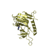
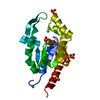
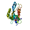
| Entry | Database: PDB / ID: 3gey | ||||||
|---|---|---|---|---|---|---|---|
| Title | Crystal structure of human poly(ADP-ribose) polymerase 15, catalytic fragment in complex with an inhibitor Pj34 | ||||||
 Components Components | Poly [ADP-ribose] polymerase 15 | ||||||
 Keywords Keywords |  TRANSFERASE / PARP / POLY(ADP-RIBOSE) POLYMERASE / BAL-3 / SGC / STRUCTURAL GENOMICS CONSORTIUM / Glycosyltransferase / NAD / Nucleus / Transcription / Transcription regulation TRANSFERASE / PARP / POLY(ADP-RIBOSE) POLYMERASE / BAL-3 / SGC / STRUCTURAL GENOMICS CONSORTIUM / Glycosyltransferase / NAD / Nucleus / Transcription / Transcription regulation | ||||||
| Function / homology |  Function and homology information Function and homology informationprotein poly-ADP-ribosylation / NAD+-protein ADP-ribosyltransferase activity / NAD+ ADP-ribosyltransferase activity / Transferases; Glycosyltransferases; Pentosyltransferases / NAD+ binding / nucleotidyltransferase activity / transcription corepressor activity / negative regulation of gene expression / negative regulation of transcription by RNA polymerase II / nucleus / cytoplasmSimilarity search - Function | ||||||
| Biological species |  Homo sapiens (human) Homo sapiens (human) | ||||||
| Method | X-RAY DIFFRACTION / SYNCHROTRON / MOLECULAR REPLACEMENT / Resolution: 2.2 Å | ||||||
 Authors Authors | Karlberg, T. / Siponen, M.I. / Arrowsmith, C.H. / Berglund, H. / Bountra, C. / Collins, R. / Edwards, A.M. / Flodin, S. / Flores, A. / Graslund, S. ...Karlberg, T. / Siponen, M.I. / Arrowsmith, C.H. / Berglund, H. / Bountra, C. / Collins, R. / Edwards, A.M. / Flodin, S. / Flores, A. / Graslund, S. / Hammarstrom, M. / Johansson, A. / Johansson, I. / Kotenyova, T. / Moche, M. / Nordlund, P. / Nyman, T. / Persson, C. / Sagemark, J. / Schutz, P. / Thorsell, A.G. / Tresaugues, L. / Van Den Berg, S. / Weigelt, J. / Welin, M. / Wisniewska, M. / Schuler, H. / Structural Genomics Consortium (SGC) | ||||||
 Citation Citation | Journal: J.Biol.Chem. / Year: 2015 Title: Structural Basis for Lack of ADP-ribosyltransferase Activity in Poly(ADP-ribose) Polymerase-13/Zinc Finger Antiviral Protein. Authors: Karlberg, T. / Klepsch, M. / Thorsell, A.G. / Andersson, C.D. / Linusson, A. / Schuler, H. | ||||||
| History |
|
- Structure visualization
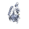
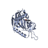
| Structure viewer | Molecule: MolmilJmol/JSmol |
|---|
- Downloads & links
-Download
| PDBx/mmCIF format | 3gey.cif.gz | 163.8 KB | Display | PDBx/mmCIF format |
|---|---|---|---|---|
| PDB format | pdb3gey.ent.gz | 130.2 KB | Display | PDB format |
| PDBx/mmJSON format | 3gey.json.gz | Tree view | PDBx/mmJSON format | |
| Others |  Other downloads Other downloads |
-Validation report
| Arichive directory | https://data.pdbj.org/pub/pdb/validation_reports/ge/3geyftp://data.pdbj.org/pub/pdb/validation_reports/ge/3gey | HTTPS FTP |
|---|
-Related structure data
| Related structure data |  2pqfC  2x5yC 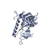 3bljSC  4x52C S: Starting model for refinement C: citing same article ( |
|---|---|
| Similar structure data |


-Links
 PDBj
PDBj- Assembly
| Deposited unit | 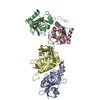
| ||||||||
|---|---|---|---|---|---|---|---|---|---|
| 1 | 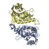
| ||||||||
| 2 | 
| ||||||||
| Unit cell |
|
-Components
| #1: Protein | Mass: 25439.459 Da / Num. of mol.: 4 / Fragment: Catalytic domain: Residues 459-656 Source method: isolated from a genetically manipulated source Source: (gene. exp.) Homo sapiens (human) / Gene: BAL3, PARP15 / Plasmid: pNIC-Bsa4 / Production host:  Escherichia coli (E. coli) / Strain (production host): BL21(DE3) R3 pRARE / References: UniProt: Q460N3, NAD+ ADP-ribosyltransferase Escherichia coli (E. coli) / Strain (production host): BL21(DE3) R3 pRARE / References: UniProt: Q460N3, NAD+ ADP-ribosyltransferase#2: Chemical |   Mass: 295.336 Da / Num. of mol.: 2 / Source method: obtained synthetically / Formula: C17H17N3O2 Mass: 295.336 Da / Num. of mol.: 2 / Source method: obtained synthetically / Formula: C17H17N3O2#3: Water | ChemComp-HOH / | Water Mass: 18.015 Da / Num. of mol.: 27 / Source method: isolated from a natural source / Formula: H2O Mass: 18.015 Da / Num. of mol.: 27 / Source method: isolated from a natural source / Formula: H2O |
|---|
-Experimental details
-Experiment
| Experiment | Method: X-RAY DIFFRACTION / Number of used crystals: 1 |
|---|
- Sample preparation
| Crystal | Density Matthews: 2.07 Å3/Da / Density % sol: 40.71 % |
|---|---|
| Crystal grow | Temperature: 293 K / Method: vapor diffusion, sitting drop / pH: 7.5 Details: 26% PEG 3350, 0.1M HEPES, pH 7.5, VAPOR DIFFUSION, SITTING DROP, temperature 293K |
-Data collection
| Diffraction | Mean temperature: 100 K |
|---|---|
| Diffraction source | Source: SYNCHROTRON / Site: Diamond  / Beamline: I03 / Wavelength: 0.98004 Å / Beamline: I03 / Wavelength: 0.98004 Å |
| Detector | Type: ADSC QUANTUM Q315r / Detector: CCD / Date: Dec 10, 2008 Details: Kirkpatrick Baez bimorph mirror pair for horizontal and vertical focusing |
| Radiation | Monochromator: double crystal / Protocol: SINGLE WAVELENGTH / Monochromatic (M) / Laue (L): M / Scattering type: x-ray |
| Radiation wavelength | Wavelength: 0.98004 Å / Relative weight: 1 |
| Reflection | Resolution: 2.2→25 Å / Num. all: 42012 / Num. obs: 42012 / % possible obs: 99.3 % / Observed criterion σ(F): 0 / Observed criterion σ(I): 0 / Redundancy: 3.7 % / Rmerge(I) obs: 0.102 / Rsym value: 0.097 / Net I/σ(I): 10.4 |
| Reflection shell | Resolution: 2.2→2.26 Å / Redundancy: 3.5 % / Rmerge(I) obs: 0.224 / Mean I/σ(I) obs: 5.7 / Rsym value: 0.256 / % possible all: 98.7 |
- Processing
| Software |
| |||||||||||||||||||||||||||||||||||||||||||||||||||||||||||||||||||||||||||||||||||||||||||||||||||||||||||||||||||||||||||||||||||||||||||||||||||
|---|---|---|---|---|---|---|---|---|---|---|---|---|---|---|---|---|---|---|---|---|---|---|---|---|---|---|---|---|---|---|---|---|---|---|---|---|---|---|---|---|---|---|---|---|---|---|---|---|---|---|---|---|---|---|---|---|---|---|---|---|---|---|---|---|---|---|---|---|---|---|---|---|---|---|---|---|---|---|---|---|---|---|---|---|---|---|---|---|---|---|---|---|---|---|---|---|---|---|---|---|---|---|---|---|---|---|---|---|---|---|---|---|---|---|---|---|---|---|---|---|---|---|---|---|---|---|---|---|---|---|---|---|---|---|---|---|---|---|---|---|---|---|---|---|---|---|---|---|
| Refinement | Method to determine structure: MOLECULAR REPLACEMENT Starting model: PDB entry 3BLJ Resolution: 2.2→24.193 Å / σ(F): 1.99 / Phase error: 38.48 / Stereochemistry target values: TWIN_LSQ_F
| |||||||||||||||||||||||||||||||||||||||||||||||||||||||||||||||||||||||||||||||||||||||||||||||||||||||||||||||||||||||||||||||||||||||||||||||||||
| Solvent computation | Shrinkage radii: 0.9 Å / VDW probe radii: 1.11 Å / Solvent model: FLAT BULK SOLVENT MODEL / Bsol: 26.866 Å2 / ksol: 0.397 e/Å3 | |||||||||||||||||||||||||||||||||||||||||||||||||||||||||||||||||||||||||||||||||||||||||||||||||||||||||||||||||||||||||||||||||||||||||||||||||||
| Refinement step | Cycle: LAST / Resolution: 2.2→24.193 Å
| |||||||||||||||||||||||||||||||||||||||||||||||||||||||||||||||||||||||||||||||||||||||||||||||||||||||||||||||||||||||||||||||||||||||||||||||||||
| Refine LS restraints |
| |||||||||||||||||||||||||||||||||||||||||||||||||||||||||||||||||||||||||||||||||||||||||||||||||||||||||||||||||||||||||||||||||||||||||||||||||||
| LS refinement shell |
|
