 Movie
Movie Controller

[English] 日本語
 Yorodumi
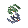
- PDB-3gec: Crystal structure of a tandem PAS domain fragment of Drosophila PERIOD -
+ Open data

- Basic information
| Entry | Database: PDB / ID: 3gec | ||||||
|---|---|---|---|---|---|---|---|
| Title | Crystal structure of a tandem PAS domain fragment of Drosophila PERIOD | ||||||
 Components Components | Period circadian protein | ||||||
 Keywords Keywords |  CIRCADIAN CLOCK PROTEIN / monomeric PAS repeat fragment / Alternative splicing / Biological rhythms / Cytoplasm / Nucleus / Phosphoprotein / Polymorphism CIRCADIAN CLOCK PROTEIN / monomeric PAS repeat fragment / Alternative splicing / Biological rhythms / Cytoplasm / Nucleus / Phosphoprotein / Polymorphism | ||||||
| Function / homology |  Function and homology information Function and homology informationNuclear import of PER and TIM / Dephosphorylation of TIM / eclosion rhythm / Transcription repression by PER and activation by PDP1 / Dephosphorylation of PER / Phosphorylation of PER and TIM / copulation / Degradation of PER / Degradation of TIM / courtship behavior ...Nuclear import of PER and TIM / Dephosphorylation of TIM / eclosion rhythm / Transcription repression by PER and activation by PDP1 / Dephosphorylation of PER / Phosphorylation of PER and TIM / copulation / Degradation of PER / Degradation of TIM / courtship behavior / male courtship behavior, veined wing generated song production / circadian temperature homeostasis / rhythmic behavior / circadian sleep/wake cycle / regulation of locomotor rhythm / regulation of circadian sleep/wake cycle, sleep / entrainment of circadian clock / mating behavior / circadian behavior / response to temperature stimulus / entrainment of circadian clock by photoperiod / locomotor rhythm / behavioral response to cocaine / response to light stimulus / long-term memory / transcription corepressor binding / determination of adult lifespan / transcription coregulator activity / regulation of protein phosphorylation / circadian regulation of gene expression / regulation of circadian rhythm / circadian rhythm / transcription corepressor activity / cell body / response to oxidative stress / transcription cis-regulatory region binding / negative regulation of DNA-templated transcription / perinuclear region of cytoplasm / negative regulation of transcription by RNA polymerase II / nucleoplasm / identical protein binding / nucleus / cytosol / cytoplasmSimilarity search - Function | ||||||
| Biological species |  Drosophila melanogaster (fruit fly) Drosophila melanogaster (fruit fly) | ||||||
| Method | X-RAY DIFFRACTION / SYNCHROTRON / MOLECULAR REPLACEMENT / Resolution: 4 Å | ||||||
 Authors Authors | Yildiz, O. / Wolf, E. | ||||||
 Citation Citation | Journal: Plos Biol. / Year: 2009 Title: Structural and functional analyses of PAS domain interactions of the clock proteins Drosophila PERIOD and mouse PERIOD2 Authors: Hennig, S. / Strauss, H.M. / Vanselow, K. / Yildiz, O. / Schulze, S. / Arens, J. / Kramer, A. / Wolf, E. | ||||||
| History |
|
- Structure visualization
| Structure viewer | Molecule: MolmilJmol/JSmol |
|---|
- Downloads & links
-Download
| PDBx/mmCIF format | 3gec.cif.gz | 61 KB | Display | PDBx/mmCIF format |
|---|---|---|---|---|
| PDB format | pdb3gec.ent.gz | 44.2 KB | Display | PDB format |
| PDBx/mmJSON format | 3gec.json.gz | Tree view | PDBx/mmJSON format | |
| Others |  Other downloads Other downloads |
-Validation report
| Arichive directory | https://data.pdbj.org/pub/pdb/validation_reports/ge/3gecftp://data.pdbj.org/pub/pdb/validation_reports/ge/3gec | HTTPS FTP |
|---|
-Related structure data
| Related structure data |  3gdiC 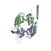 1wa9S S: Starting model for refinement C: citing same article ( |
|---|---|
| Similar structure data |

-Links
 PDBj
PDBj


- Assembly
| Deposited unit | 
| ||||||||
|---|---|---|---|---|---|---|---|---|---|
| 1 |
| ||||||||
| Unit cell |
|
-Components
| #1: Protein | Mass: 34940.168 Da / Num. of mol.: 1 / Fragment: PAS domain Source method: isolated from a genetically manipulated source Source: (gene. exp.) Drosophila melanogaster (fruit fly) / Gene: per / Plasmid: pGEX6P2 / Production host:  Escherichia coli BL21(DE3) (bacteria) / Strain (production host): BL21(DE3) / References: UniProt: P07663 Escherichia coli BL21(DE3) (bacteria) / Strain (production host): BL21(DE3) / References: UniProt: P07663 |
|---|
-Experimental details
-Experiment
| Experiment | Method: X-RAY DIFFRACTION / Number of used crystals: 1 |
|---|
- Sample preparation
| Crystal | Density Matthews: 4.68 Å3/Da / Density % sol: 73.71 % |
|---|---|
| Crystal grow | Temperature: 282 K / Method: vapor diffusion, hanging drop / pH: 7.7 Details: 70mM Sodiumtartrate, 20mM Hepes, 5mM DTE, pH 7.7, VAPOR DIFFUSION, HANGING DROP, temperature 282K |
-Data collection
| Diffraction | Mean temperature: 100 K |
|---|---|
| Diffraction source | Source: SYNCHROTRON / Site: ESRF  / Beamline: ID29 / Wavelength: 0.9793 Å / Beamline: ID29 / Wavelength: 0.9793 Å |
| Detector | Type: ADSC QUANTUM 4 / Detector: CCD |
| Radiation | Protocol: SINGLE WAVELENGTH / Monochromatic (M) / Laue (L): M / Scattering type: x-ray |
| Radiation wavelength | Wavelength: 0.9793 Å / Relative weight: 1 |
| Reflection | Resolution: 4→19.68 Å / Num. obs: 5578 / % possible obs: 96.7 % / Observed criterion σ(F): 0 / Observed criterion σ(I): -3 / Redundancy: 5.4 % / Biso Wilson estimate: 136.8 Å2 / Rmerge(I) obs: 0.055 / Rsym value: 0.061 / Net I/σ(I): 18.07 |
| Reflection shell | Resolution: 4→4.1 Å / Redundancy: 5.6 % / Rmerge(I) obs: 0.422 / Mean I/σ(I) obs: 3.82 / Num. unique all: 408 / Rsym value: 0.469 / % possible all: 97.8 |
- Processing
| Software |
| |||||||||||||||||||||||||||||||||||||||||||||||||
|---|---|---|---|---|---|---|---|---|---|---|---|---|---|---|---|---|---|---|---|---|---|---|---|---|---|---|---|---|---|---|---|---|---|---|---|---|---|---|---|---|---|---|---|---|---|---|---|---|---|---|
| Refinement | Method to determine structure: MOLECULAR REPLACEMENT Starting model: 1WA9 Resolution: 4→19.68 Å / Cross valid method: THROUGHOUT / σ(F): 0 / σ(I): 0 / Stereochemistry target values: Engh & Huber
| |||||||||||||||||||||||||||||||||||||||||||||||||
| Displacement parameters | Biso mean: 151.3 Å2
| |||||||||||||||||||||||||||||||||||||||||||||||||
| Refine analyze |
| |||||||||||||||||||||||||||||||||||||||||||||||||
| Refinement step | Cycle: LAST / Resolution: 4→19.68 Å
| |||||||||||||||||||||||||||||||||||||||||||||||||
| Refine LS restraints |
| |||||||||||||||||||||||||||||||||||||||||||||||||
| LS refinement shell | Refine-ID: X-RAY DIFFRACTION / Total num. of bins used: 6
|
