 Movie
Movie Controller

[English] 日本語
 Yorodumi
- PDB-1wa9: Crystal Structure of the PAS repeat region of the Drosophila cloc... -
+ Open data

- Basic information
| Entry | Database: PDB / ID: 1wa9 | ||||||
|---|---|---|---|---|---|---|---|
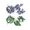
| Title | Crystal Structure of the PAS repeat region of the Drosophila clock protein PERIOD | ||||||
 Components Components | PERIOD CIRCADIAN PROTEIN | ||||||
 Keywords Keywords |  CIRCADIAN RHYTHM / PERIOD / PAS DOMAIN / CLOCK PROTEIN / PHOSPHORYLATION / POLYMORPHISM CIRCADIAN RHYTHM / PERIOD / PAS DOMAIN / CLOCK PROTEIN / PHOSPHORYLATION / POLYMORPHISM | ||||||
| Function / homology |  Function and homology information Function and homology informationNuclear import of PER and TIM / Dephosphorylation of TIM / eclosion rhythm / Transcription repression by PER and activation by PDP1 / Dephosphorylation of PER / Phosphorylation of PER and TIM / copulation / Degradation of PER / Degradation of TIM / courtship behavior ...Nuclear import of PER and TIM / Dephosphorylation of TIM / eclosion rhythm / Transcription repression by PER and activation by PDP1 / Dephosphorylation of PER / Phosphorylation of PER and TIM / copulation / Degradation of PER / Degradation of TIM / courtship behavior / male courtship behavior, veined wing generated song production / circadian temperature homeostasis / rhythmic behavior / circadian sleep/wake cycle / regulation of locomotor rhythm / regulation of circadian sleep/wake cycle, sleep / entrainment of circadian clock / mating behavior / circadian behavior / response to temperature stimulus / entrainment of circadian clock by photoperiod / locomotor rhythm / behavioral response to cocaine / response to light stimulus / long-term memory / transcription corepressor binding / determination of adult lifespan / transcription coregulator activity / regulation of protein phosphorylation / circadian regulation of gene expression / regulation of circadian rhythm / circadian rhythm / transcription corepressor activity / cell body / response to oxidative stress / transcription cis-regulatory region binding / negative regulation of DNA-templated transcription / perinuclear region of cytoplasm / negative regulation of transcription by RNA polymerase II / nucleoplasm / identical protein binding / nucleus / cytosol / cytoplasmSimilarity search - Function | ||||||
| Biological species |  DROSOPHILA MELANOGASTER (fruit fly) DROSOPHILA MELANOGASTER (fruit fly) | ||||||
| Method | X-RAY DIFFRACTION / SYNCHROTRON / MAD / Resolution: 3.15 Å | ||||||
 Authors Authors | Yildiz, O. / Doi, M. / Yujnovsky, I. / Cardone, L. / Berndt, A. / Hennig, S. / Schulze, S. / Urbanke, C. / Sassone-Corsi, P. / Wolf, E. | ||||||
 Citation Citation | Journal: Mol.Cell / Year: 2005 Title: Crystal Structure and Interactions of the Pas Repeat Region of the Drosophila Clock Protein Period Authors: Yildiz, O. / Doi, M. / Yujnovsky, I. / Cardone, L. / Berndt, A. / Hennig, S. / Schulze, S. / Urbanke, C. / Sassone-Corsi, P. / Wolf, E. | ||||||
| History |
| ||||||
| Remark 700 | SHEET THE SHEET STRUCTURE OF THIS MOLECULE IS BIFURCATED. IN ORDER TO REPRESENT THIS FEATURE IN ... SHEET THE SHEET STRUCTURE OF THIS MOLECULE IS BIFURCATED. IN ORDER TO REPRESENT THIS FEATURE IN THE SHEET RECORDS BELOW, TWO SHEETS ARE DEFINED. |
- Structure visualization
| Structure viewer | Molecule: MolmilJmol/JSmol |
|---|
- Downloads & links
-Download
| PDBx/mmCIF format | 1wa9.cif.gz | 125.5 KB | Display | PDBx/mmCIF format |
|---|---|---|---|---|
| PDB format | pdb1wa9.ent.gz | 99.5 KB | Display | PDB format |
| PDBx/mmJSON format | 1wa9.json.gz | Tree view | PDBx/mmJSON format | |
| Others |  Other downloads Other downloads |
-Validation report
| Arichive directory | https://data.pdbj.org/pub/pdb/validation_reports/wa/1wa9ftp://data.pdbj.org/pub/pdb/validation_reports/wa/1wa9 | HTTPS FTP |
|---|
-Related structure data
| Similar structure data |
|---|


-Links
 PDBj
PDBj


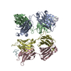
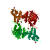
- Assembly
| Deposited unit | 
| ||||||||
|---|---|---|---|---|---|---|---|---|---|
| 1 |
| ||||||||
| Unit cell |
| ||||||||
| Noncrystallographic symmetry (NCS) | NCS oper: (Code: given Matrix: (-0.9529, 0.0677, -0.2955), Vector : |
-Components
| #1: Protein | Mass: 41457.469 Da / Num. of mol.: 2 / Fragment: PAS REPEAT REGION, RESIDUES 232-599 Source method: isolated from a genetically manipulated source Source: (gene. exp.) DROSOPHILA MELANOGASTER (fruit fly) / Production host:  ESCHERICHIA COLI (E. coli) / Strain (production host): BL21(DE3) / References: UniProt: P07663 ESCHERICHIA COLI (E. coli) / Strain (production host): BL21(DE3) / References: UniProt: P07663#2: Water | ChemComp-HOH / | Water Mass: 18.015 Da / Num. of mol.: 6 / Source method: isolated from a natural source / Formula: H2O Mass: 18.015 Da / Num. of mol.: 6 / Source method: isolated from a natural source / Formula: H2O |
|---|
-Experimental details
-Experiment
| Experiment | Method: X-RAY DIFFRACTION / Number of used crystals: 1 |
|---|
- Sample preparation
| Crystal | Density Matthews: 3.7 Å3/Da / Density % sol: 65 % |
|---|---|
| Crystal grow | pH: 7.7 / Details: 500 MM CALCIUM CHLORIDE, pH 7.70 |
-Data collection
| Diffraction | Mean temperature: 100 K |
|---|---|
| Diffraction source | Source: SYNCHROTRON / Site: ESRF  / Beamline: ID14-2 / Wavelength: 0.933 / Beamline: ID14-2 / Wavelength: 0.933 |
| Detector | Type: ADSC CCD / Detector: CCD / Date: Jul 19, 2002 / Details: TOROIDAL MIRROR |
| Radiation | Monochromator: DIAMOND CRYSTAL / Protocol: SINGLE WAVELENGTH / Monochromatic (M) / Laue (L): M / Scattering type: x-ray |
| Radiation wavelength | Wavelength: 0.933 Å / Relative weight: 1 |
| Reflection | Resolution: 3.15→25 Å / Num. obs: 21048 / % possible obs: 99.8 % / Observed criterion σ(I): 0 / Redundancy: 4.9 % / Biso Wilson estimate: 87.6 Å2 / Rmerge(I) obs: 0.05 / Rsym value: 0.06 / Net I/σ(I): 18.1 |
| Reflection shell | Resolution: 3.15→3.25 Å / Redundancy: 5 % / Rmerge(I) obs: 0.35 / Mean I/σ(I) obs: 4.6 / Rsym value: 0.38 / % possible all: 99.9 |
- Processing
| Software |
| ||||||||||||||||||||||||||||||||||||||||||||||||||||||||||||
|---|---|---|---|---|---|---|---|---|---|---|---|---|---|---|---|---|---|---|---|---|---|---|---|---|---|---|---|---|---|---|---|---|---|---|---|---|---|---|---|---|---|---|---|---|---|---|---|---|---|---|---|---|---|---|---|---|---|---|---|---|---|
| Refinement | Method to determine structure: MAD / Resolution: 3.15→25 Å / Rfactor Rfree error: 0.009 / Data cutoff high absF: 10000 / Cross valid method: THROUGHOUT / σ(F): 0 / Stereochemistry target values: MAXIMUM LIKELIHOOD
| ||||||||||||||||||||||||||||||||||||||||||||||||||||||||||||
| Solvent computation | Solvent model: FLAT MODEL / Bsol: 58.4308 Å2 / ksol: 0.213266 e/Å3 | ||||||||||||||||||||||||||||||||||||||||||||||||||||||||||||
| Displacement parameters | Biso mean: 63.9 Å2
| ||||||||||||||||||||||||||||||||||||||||||||||||||||||||||||
| Refine analyze |
| ||||||||||||||||||||||||||||||||||||||||||||||||||||||||||||
| Refinement step | Cycle: LAST / Resolution: 3.15→25 Å
| ||||||||||||||||||||||||||||||||||||||||||||||||||||||||||||
| Refine LS restraints |
| ||||||||||||||||||||||||||||||||||||||||||||||||||||||||||||
| LS refinement shell | Resolution: 3.15→3.26 Å / Rfactor Rfree error: 0.051 / Total num. of bins used: 10
| ||||||||||||||||||||||||||||||||||||||||||||||||||||||||||||
| Xplor file |
|
