 Movie
Movie Controller


+ Open data

- Basic information
| Entry | Database: PDB / ID: 3g7l | ||||||
|---|---|---|---|---|---|---|---|
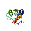
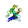
| Title | Chromodomain of Chp1 in complex with Histone H3K9me3 peptide | ||||||
 Components Components |
| ||||||
 Keywords Keywords |  NUCLEAR PROTEIN / chromodomain / protein-peptide complex / silencing / Cell cycle / Chromosome partition / DNA-binding / Nucleus / RNA-mediated gene silencing / Acetylation / Chromosomal protein / DNA damage / DNA repair / Methylation / Nucleosome core / Phosphoprotein NUCLEAR PROTEIN / chromodomain / protein-peptide complex / silencing / Cell cycle / Chromosome partition / DNA-binding / Nucleus / RNA-mediated gene silencing / Acetylation / Chromosomal protein / DNA damage / DNA repair / Methylation / Nucleosome core / Phosphoprotein | ||||||
| Function / homology |  Function and homology information Function and homology informationnucleolar peripheral inclusion body / RITS complex / siRNA-mediated pericentric heterochromatin formation / mating-type region heterochromatin / heterochromatin island / heterochromatin boundary formation / mitotic sister chromatid biorientation / regulatory ncRNA-mediated heterochromatin formation / chromosome, subtelomeric region / chromatin-protein adaptor activity ...nucleolar peripheral inclusion body / RITS complex / siRNA-mediated pericentric heterochromatin formation / mating-type region heterochromatin / heterochromatin island / heterochromatin boundary formation / mitotic sister chromatid biorientation / regulatory ncRNA-mediated heterochromatin formation / chromosome, subtelomeric region / chromatin-protein adaptor activity / spindle pole body / subtelomeric heterochromatin formation / heterochromatin / pericentric heterochromatin / methylated histone binding / histone reader activity / chromosome segregation / structural constituent of chromatin / nucleosome / chromatin organization / histone binding / single-stranded RNA binding / protein heterodimerization activity / DNA damage response / chromatin binding / negative regulation of transcription by RNA polymerase II / DNA binding / nucleus / cytoplasmSimilarity search - Function | ||||||
| Biological species |  Schizosaccharomyces pombe (fission yeast) Schizosaccharomyces pombe (fission yeast) | ||||||
| Method | X-RAY DIFFRACTION / SYNCHROTRON / MOLECULAR REPLACEMENT / Resolution: 2.2 Å | ||||||
 Authors Authors | Schalch, T. / Joshua-Tor, L. | ||||||
 Citation Citation | Journal: Mol.Cell / Year: 2009 Title: High-affinity binding of Chp1 chromodomain to K9 methylated histone H3 is required to establish centromeric heterochromatin Authors: Schalch, T. / Job, G. / Noffsinger, V.J. / Shanker, S. / Kuscu, C. / Joshua-Tor, L. / Partridge, J.F. | ||||||
| History |
|
- Structure visualization
| Structure viewer | Molecule: MolmilJmol/JSmol |
|---|
- Downloads & links
-Download
| PDBx/mmCIF format | 3g7l.cif.gz | 54.8 KB | Display | PDBx/mmCIF format |
|---|---|---|---|---|
| PDB format | pdb3g7l.ent.gz | 39.8 KB | Display | PDB format |
| PDBx/mmJSON format | 3g7l.json.gz | Tree view | PDBx/mmJSON format | |
| Others |  Other downloads Other downloads |
-Validation report
| Arichive directory | https://data.pdbj.org/pub/pdb/validation_reports/g7/3g7lftp://data.pdbj.org/pub/pdb/validation_reports/g7/3g7l | HTTPS FTP |
|---|
-Related structure data
| Related structure data |  1kneS S: Starting model for refinement |
|---|---|
| Similar structure data |
-Links
 PDBj
PDBj


- Assembly
| Deposited unit | 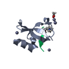
| ||||||||
|---|---|---|---|---|---|---|---|---|---|
| 1 |
| ||||||||
| Unit cell |
|
-Components
| #1: Protein | Mass: 7383.179 Da / Num. of mol.: 1 / Fragment: Chromodomain (UNP residues 15 to 75) Source method: isolated from a genetically manipulated source Details: N-terminal His6-Sumo fusion Source: (gene. exp.) Schizosaccharomyces pombe (fission yeast)Gene: chp1, SPAC18G6.02c / Plasmid: pET28a-SUMO / Production host:  Escherichia coli (E. coli) / Strain (production host): BL21 (DE3) RIPL / References: UniProt: Q10103 Escherichia coli (E. coli) / Strain (production host): BL21 (DE3) RIPL / References: UniProt: Q10103 | ||
|---|---|---|---|
| #2: Protein/peptide | Mass: 1771.051 Da / Num. of mol.: 1 / Fragment: UNP residues 2 to 17 / Mutation: R17Y / Source method: obtained synthetically Details: Peptide synthesis by Protein Peptide Research Ldt., Hampshire UK. References: UniProt: P09988 | ||
| #3: Chemical | ChemComp-ACY / Acetic acid  Mass: 60.052 Da / Num. of mol.: 1 / Source method: obtained synthetically / Formula: C2H4O2 Mass: 60.052 Da / Num. of mol.: 1 / Source method: obtained synthetically / Formula: C2H4O2 | ||
| #4: Chemical | ChemComp-ZN /   Mass: 65.409 Da / Num. of mol.: 4 / Source method: obtained synthetically / Formula: Zn Mass: 65.409 Da / Num. of mol.: 4 / Source method: obtained synthetically / Formula: Zn#5: Water | ChemComp-HOH / | Water Mass: 18.015 Da / Num. of mol.: 29 / Source method: isolated from a natural source / Formula: H2O Mass: 18.015 Da / Num. of mol.: 29 / Source method: isolated from a natural source / Formula: H2O |
-Experimental details
-Experiment
| Experiment | Method: X-RAY DIFFRACTION / Number of used crystals: 1 |
|---|
- Sample preparation
| Crystal | Density Matthews: 2.04 Å3/Da / Density % sol: 39.68 % |
|---|---|
| Crystal grow | Temperature: 290 K / Method: vapor diffusion, hanging drop / pH: 6 Details: 0.05 M Zinc Acetate, 20% w/v PEG 3350, pH 6, VAPOR DIFFUSION, HANGING DROP, temperature 290K |
-Data collection
| Diffraction | Mean temperature: 100 K |
|---|---|
| Diffraction source | Source: SYNCHROTRON / Site: NSLS  / Beamline: X29A / Wavelength: 1.0809 Å / Beamline: X29A / Wavelength: 1.0809 Å |
| Detector | Type: ADSC QUANTUM Q315r / Detector: CCD / Date: Feb 9, 2007 |
| Radiation | Monochromator: CRYOGENICALLY COOLED DOUBLE CRYSTAL MONOCHROMATOR WITH HORIZONTAL FOCUSING SAGITTAL BEND SECOND MONO CRYSTAL WITH 4:1 MAGNIFICATION RATIO AND VERTICALLY FOCUSING MIRROR Protocol: SINGLE WAVELENGTH / Monochromatic (M) / Laue (L): M / Scattering type: x-ray |
| Radiation wavelength | Wavelength: 1.0809 Å / Relative weight: 1 |
| Reflection | Resolution: 2.2→19.3 Å / Num. all: 7418 / Num. obs: 7255 / % possible obs: 97.93 % / Observed criterion σ(F): -3 / Observed criterion σ(I): -3 / Redundancy: 3.4 % / Biso Wilson estimate: 44.94 Å2 / Rmerge(I) obs: 0.047 / Net I/σ(I): 14.87 |
| Reflection shell | Resolution: 2.2→2.4 Å / Redundancy: 3.3 % / Rmerge(I) obs: 0.314 / Mean I/σ(I) obs: 3.46 / Num. unique all: 1683 / % possible all: 99.8 |
- Processing
| Software |
| ||||||||||||||||||||||||||||||||||||||||||
|---|---|---|---|---|---|---|---|---|---|---|---|---|---|---|---|---|---|---|---|---|---|---|---|---|---|---|---|---|---|---|---|---|---|---|---|---|---|---|---|---|---|---|---|
| Refinement | Method to determine structure: MOLECULAR REPLACEMENT Starting model: Chain A of pdb code 1KNE Resolution: 2.2→19.337 Å / SU ML: 0.28 / Isotropic thermal model: Isotropic and TLS / Cross valid method: THROUGHOUT / σ(F): 0 / Stereochemistry target values: CCP4 monomer library
| ||||||||||||||||||||||||||||||||||||||||||
| Solvent computation | Shrinkage radii: 0.9 Å / VDW probe radii: 1.11 Å / Solvent model: FLAT BULK SOLVENT MODEL / Bsol: 56.891 Å2 / ksol: 0.41 e/Å3 | ||||||||||||||||||||||||||||||||||||||||||
| Displacement parameters | Biso mean: 38.5 Å2 | ||||||||||||||||||||||||||||||||||||||||||
| Refinement step | Cycle: LAST / Resolution: 2.2→19.337 Å
| ||||||||||||||||||||||||||||||||||||||||||
| Refine LS restraints |
| ||||||||||||||||||||||||||||||||||||||||||
| LS refinement shell |
| ||||||||||||||||||||||||||||||||||||||||||
| Refinement TLS params. | Method: refined / Origin x: 0.5302 Å / Origin y: -17.8348 Å / Origin z: -9.4336 Å
| ||||||||||||||||||||||||||||||||||||||||||
| Refinement TLS group | Selection details: all |
