 Movie
Movie Controller

[English] 日本語
 Yorodumi
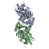
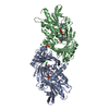
- PDB-3g3e: Crystal structure of human D-amino acid oxidase in complex with h... -
+ Open data

- Basic information
| Entry | Database: PDB / ID: 3g3e | ||||||
|---|---|---|---|---|---|---|---|
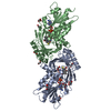
| Title | Crystal structure of human D-amino acid oxidase in complex with hydroxyquinolin-2(1H) | ||||||
 Components Components | D-amino-acid oxidase D-amino acid oxidase D-amino acid oxidase | ||||||
 Keywords Keywords | OXIDOREDUCTASE / d-amino acid oxidase / FAD / Flavoprotein / Peroxisome | ||||||
| Function / homology |  Function and homology information Function and homology informationD-alanine catabolic process / D-amino-acid oxidase / D-amino-acid oxidase activity / D-serine metabolic process / D-serine catabolic process / proline catabolic process / D-amino acid catabolic process / Glyoxylate metabolism and glycine degradation / dopamine biosynthetic process / presynaptic active zone ...D-alanine catabolic process / D-amino-acid oxidase / D-amino-acid oxidase activity / D-serine metabolic process / D-serine catabolic process / proline catabolic process / D-amino acid catabolic process / Glyoxylate metabolism and glycine degradation / dopamine biosynthetic process / presynaptic active zone / neutrophil-mediated killing of gram-negative bacterium / peroxisomal matrix / digestion / FAD binding / Peroxisomal protein import / identical protein binding / cytosol / cytoplasmSimilarity search - Function | ||||||
| Biological species |  Homo sapiens (human) Homo sapiens (human) | ||||||
| Method | X-RAY DIFFRACTION / SYNCHROTRON / MOLECULAR REPLACEMENT / Resolution: 2.2 Å | ||||||
 Authors Authors | Duplantier, A. / Liu, S. | ||||||
 Citation Citation | Journal: J.Med.Chem. / Year: 2009 Title: Discovery, SAR, and pharmacokinetics of a novel 3-Hydroxyquinolin-2(1H)-one series of potent D-amino acid oxidase (DAAO) inhibitors Authors: Duplantier, A.J. / Becker, S.L. / Bohanon, M.J. / Borzilleri, K.A. / Chrunyk, B.A. / Downs, J.T. / Hu, L.Y. / El-Kattan, A. / James, L.C. / Liu, S. / Lu, J. / Maklad, N. / Mansour, M.N. / ...Authors: Duplantier, A.J. / Becker, S.L. / Bohanon, M.J. / Borzilleri, K.A. / Chrunyk, B.A. / Downs, J.T. / Hu, L.Y. / El-Kattan, A. / James, L.C. / Liu, S. / Lu, J. / Maklad, N. / Mansour, M.N. / Mente, S. / Piotrowski, M.A. / Sakya, S.M. / Sheehan, S. / Steyn, S.J. / Strick, C.A. / Williams, V.A. / Zhang, L. | ||||||
| History |
|
- Structure visualization
| Structure viewer | Molecule: MolmilJmol/JSmol |
|---|
- Downloads & links
-Download
| PDBx/mmCIF format | 3g3e.cif.gz | 291.7 KB | Display | PDBx/mmCIF format |
|---|---|---|---|---|
| PDB format | pdb3g3e.ent.gz | 237.5 KB | Display | PDB format |
| PDBx/mmJSON format | 3g3e.json.gz | Tree view | PDBx/mmJSON format | |
| Others |  Other downloads Other downloads |
-Validation report
| Arichive directory | https://data.pdbj.org/pub/pdb/validation_reports/g3/3g3eftp://data.pdbj.org/pub/pdb/validation_reports/g3/3g3e | HTTPS FTP |
|---|
-Related structure data
| Related structure data | 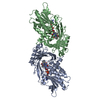 1an9S S: Starting model for refinement |
|---|---|
| Similar structure data |

-Links
 PDBj
PDBj- Assembly
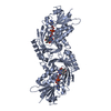
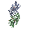
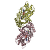
| Deposited unit | 
| |||||||||||||||
|---|---|---|---|---|---|---|---|---|---|---|---|---|---|---|---|---|
| 1 | 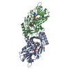
| |||||||||||||||
| 2 | 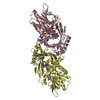
| |||||||||||||||
| Unit cell |
| |||||||||||||||
| Noncrystallographic symmetry (NCS) | NCS domain:
|
-Components
| #1: Protein | D-amino acid oxidase / DAMOX / DAAO / DAO Mass: 39987.504 Da / Num. of mol.: 4 Source method: isolated from a genetically manipulated source Source: (gene. exp.) Homo sapiens (human) / Gene: DAO, DAMOX / Production host:  Escherichia coli (E. coli) / Strain (production host): BL21 / References: UniProt: P14920, D-amino-acid oxidase Escherichia coli (E. coli) / Strain (production host): BL21 / References: UniProt: P14920, D-amino-acid oxidase#2: Chemical | ChemComp-FAD / Flavin adenine dinucleotide  Mass: 785.550 Da / Num. of mol.: 4 / Source method: obtained synthetically / Formula: C27H33N9O15P2 / Comment: FAD*YM Mass: 785.550 Da / Num. of mol.: 4 / Source method: obtained synthetically / Formula: C27H33N9O15P2 / Comment: FAD*YM#3: Chemical | ChemComp-G3E /   Mass: 161.157 Da / Num. of mol.: 4 / Source method: obtained synthetically / Formula: C9H7NO2 Mass: 161.157 Da / Num. of mol.: 4 / Source method: obtained synthetically / Formula: C9H7NO2#4: Water | ChemComp-HOH / | Water Mass: 18.015 Da / Num. of mol.: 320 / Source method: isolated from a natural source / Formula: H2O Mass: 18.015 Da / Num. of mol.: 320 / Source method: isolated from a natural source / Formula: H2O |
|---|
-Experimental details
-Experiment
| Experiment | Method: X-RAY DIFFRACTION |
|---|
- Sample preparation
| Crystal | Density Matthews: 2.16 Å3/Da / Density % sol: 43.07 % |
|---|
-Data collection
| Diffraction | Mean temperature: 100 K |
|---|---|
| Diffraction source | Source: SYNCHROTRON / Site: APS  / Beamline: 17-ID / Wavelength: 1 Å / Beamline: 17-ID / Wavelength: 1 Å |
| Detector | Type: ADSC QUANTUM 315 / Detector: CCD / Date: Jun 2, 2008 |
| Radiation | Protocol: SINGLE WAVELENGTH / Monochromatic (M) / Laue (L): M / Scattering type: x-ray |
| Radiation wavelength | Wavelength: 1 Å / Relative weight: 1 |
| Reflection | Resolution: 2.2→50 Å / Num. obs: 61073 / % possible obs: 88.5 % / Observed criterion σ(I): 2 / Redundancy: 1.7 % / Rmerge(I) obs: 0.068 |
- Processing
| Software | Name: REFMAC / Version: 5.4.0069 / Classification: refinement | ||||||||||||||||||||||||||||||||||||||||||||||||||||||||||||||||||||||||||||||||||||||||||||||||||||||||||||||||||||||||||||||||||||||||||||||||||||||||||||||||||||||||||
|---|---|---|---|---|---|---|---|---|---|---|---|---|---|---|---|---|---|---|---|---|---|---|---|---|---|---|---|---|---|---|---|---|---|---|---|---|---|---|---|---|---|---|---|---|---|---|---|---|---|---|---|---|---|---|---|---|---|---|---|---|---|---|---|---|---|---|---|---|---|---|---|---|---|---|---|---|---|---|---|---|---|---|---|---|---|---|---|---|---|---|---|---|---|---|---|---|---|---|---|---|---|---|---|---|---|---|---|---|---|---|---|---|---|---|---|---|---|---|---|---|---|---|---|---|---|---|---|---|---|---|---|---|---|---|---|---|---|---|---|---|---|---|---|---|---|---|---|---|---|---|---|---|---|---|---|---|---|---|---|---|---|---|---|---|---|---|---|---|---|---|---|
| Refinement | Method to determine structure: MOLECULAR REPLACEMENT Starting model: PDB entry 1AN9 Resolution: 2.2→10 Å / Cor.coef. Fo:Fc: 0.933 / Cor.coef. Fo:Fc free: 0.899 / SU B: 20.548 / SU ML: 0.259 / TLS residual ADP flag: LIKELY RESIDUAL / Cross valid method: THROUGHOUT / ESU R: 0.586 / ESU R Free: 0.309 / Stereochemistry target values: MAXIMUM LIKELIHOOD / Details: HYDROGENS HAVE BEEN ADDED IN THE RIDING POSITIONS
| ||||||||||||||||||||||||||||||||||||||||||||||||||||||||||||||||||||||||||||||||||||||||||||||||||||||||||||||||||||||||||||||||||||||||||||||||||||||||||||||||||||||||||
| Solvent computation | Ion probe radii: 0.8 Å / Shrinkage radii: 0.8 Å / VDW probe radii: 1.4 Å / Solvent model: BABINET MODEL WITH MASK | ||||||||||||||||||||||||||||||||||||||||||||||||||||||||||||||||||||||||||||||||||||||||||||||||||||||||||||||||||||||||||||||||||||||||||||||||||||||||||||||||||||||||||
| Displacement parameters | Biso mean: 46.903 Å2
| ||||||||||||||||||||||||||||||||||||||||||||||||||||||||||||||||||||||||||||||||||||||||||||||||||||||||||||||||||||||||||||||||||||||||||||||||||||||||||||||||||||||||||
| Refinement step | Cycle: LAST / Resolution: 2.2→10 Å
| ||||||||||||||||||||||||||||||||||||||||||||||||||||||||||||||||||||||||||||||||||||||||||||||||||||||||||||||||||||||||||||||||||||||||||||||||||||||||||||||||||||||||||
| Refine LS restraints |
| ||||||||||||||||||||||||||||||||||||||||||||||||||||||||||||||||||||||||||||||||||||||||||||||||||||||||||||||||||||||||||||||||||||||||||||||||||||||||||||||||||||||||||
| Refine LS restraints NCS | Dom-ID: 1 / Ens-ID: 1 / Number: 4742 / Refine-ID: X-RAY DIFFRACTION
| ||||||||||||||||||||||||||||||||||||||||||||||||||||||||||||||||||||||||||||||||||||||||||||||||||||||||||||||||||||||||||||||||||||||||||||||||||||||||||||||||||||||||||
| LS refinement shell | Resolution: 2.2→2.254 Å / Total num. of bins used: 20
| ||||||||||||||||||||||||||||||||||||||||||||||||||||||||||||||||||||||||||||||||||||||||||||||||||||||||||||||||||||||||||||||||||||||||||||||||||||||||||||||||||||||||||
| Refinement TLS params. | Method: refined / Refine-ID: X-RAY DIFFRACTION
| ||||||||||||||||||||||||||||||||||||||||||||||||||||||||||||||||||||||||||||||||||||||||||||||||||||||||||||||||||||||||||||||||||||||||||||||||||||||||||||||||||||||||||
| Refinement TLS group |
|
