 Movie
Movie Controller

[English] 日本語
 Yorodumi
- PDB-3g2o: Crystal Structure of the Glycopeptide N-methyltransferase MtfA co... -
+ Open data

- Basic information
| Entry | Database: PDB / ID: 3g2o | ||||||
|---|---|---|---|---|---|---|---|
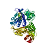
| Title | Crystal Structure of the Glycopeptide N-methyltransferase MtfA complexed with (S)-adenosyl-L-methionine (SAM) | ||||||
 Components Components | PCZA361.24 | ||||||
 Keywords Keywords |  TRANSFERASE / SAM-dependent methyltransferase / glycopeptide antibiotics biosynthesis / Structural Genomics / Montreal-Kingston Bacterial Structural Genomics Initiative / BSGI TRANSFERASE / SAM-dependent methyltransferase / glycopeptide antibiotics biosynthesis / Structural Genomics / Montreal-Kingston Bacterial Structural Genomics Initiative / BSGI | ||||||
| Function / homology |  Function and homology information Function and homology informationCarboxypeptidase Inhibitor; Chain A - #60 / HSP40/DNAj peptide-binding domain - #20 / : / HSP40/DNAj peptide-binding domain / Carboxypeptidase Inhibitor; Chain A / Methyltransferase domain 25 / Methyltransferase domain / Other non-globular / Vaccinia Virus protein VP39 / Special ...Carboxypeptidase Inhibitor; Chain A - #60 / HSP40/DNAj peptide-binding domain - #20 / : / HSP40/DNAj peptide-binding domain / Carboxypeptidase Inhibitor; Chain A / Methyltransferase domain 25 / Methyltransferase domain / Other non-globular / Vaccinia Virus protein VP39 / Special / S-adenosyl-L-methionine-dependent methyltransferase superfamily / Rossmann fold / 2-Layer Sandwich / 3-Layer(aba) Sandwich / Alpha BetaSimilarity search - Domain/homology | ||||||
| Biological species |  Amycolatopsis orientalis (bacteria) Amycolatopsis orientalis (bacteria) | ||||||
| Method | X-RAY DIFFRACTION / SYNCHROTRON / MOLECULAR REPLACEMENT / molecular replacement / Resolution: 2.1 Å | ||||||
 Authors Authors | Shi, R. / Matte, A. / Cygler, M. / Montreal-Kingston Bacterial Structural Genomics Initiative (BSGI) | ||||||
 Citation Citation | Journal: Chem.Biol. / Year: 2009 Title: Structure and function of the glycopeptide N-methyltransferase MtfA, a tool for the biosynthesis of modified glycopeptide antibiotics. Authors: Shi, R. / Lamb, S.S. / Zakeri, B. / Proteau, A. / Cui, Q. / Sulea, T. / Matte, A. / Wright, G.D. / Cygler, M. | ||||||
| History |
|
- Structure visualization
| Structure viewer | Molecule: MolmilJmol/JSmol |
|---|
- Downloads & links
-Download
| PDBx/mmCIF format | 3g2o.cif.gz | 110.5 KB | Display | PDBx/mmCIF format |
|---|---|---|---|---|
| PDB format | pdb3g2o.ent.gz | 82.8 KB | Display | PDB format |
| PDBx/mmJSON format | 3g2o.json.gz | Tree view | PDBx/mmJSON format | |
| Others |  Other downloads Other downloads |
-Validation report
| Arichive directory | https://data.pdbj.org/pub/pdb/validation_reports/g2/3g2oftp://data.pdbj.org/pub/pdb/validation_reports/g2/3g2o | HTTPS FTP |
|---|
-Related structure data
| Related structure data |  3g2mSC  3g2pC  3g2qC S: Starting model for refinement C: citing same article ( |
|---|---|
| Similar structure data | |
| Other databases |


-Links
 PDBj
PDBj
- Assembly
| Deposited unit | 
| ||||||||
|---|---|---|---|---|---|---|---|---|---|
| 1 |
| ||||||||
| Unit cell |
|
-Components
| #1: Protein | Mass: 32527.627 Da / Num. of mol.: 2 Source method: isolated from a genetically manipulated source Source: (gene. exp.) Amycolatopsis orientalis (bacteria) / Gene: mtfA / Plasmid: pET28a / Production host: Escherichia coli (E. coli) / Strain (production host): BL21(DE3) / References: UniProt: O52805#2: Chemical | S-Adenosyl methionine  Mass: 398.437 Da / Num. of mol.: 2 / Source method: obtained synthetically / Formula: C15H22N6O5S Mass: 398.437 Da / Num. of mol.: 2 / Source method: obtained synthetically / Formula: C15H22N6O5S#3: Water | ChemComp-HOH / | Water Mass: 18.015 Da / Num. of mol.: 144 / Source method: isolated from a natural source / Formula: H2O Mass: 18.015 Da / Num. of mol.: 144 / Source method: isolated from a natural source / Formula: H2O |
|---|
-Experimental details
-Experiment
| Experiment | Method: X-RAY DIFFRACTION / Number of used crystals: 1 |
|---|
- Sample preparation
| Crystal | Density Matthews: 2.58 Å3/Da / Density % sol: 52.31 % |
|---|---|
| Crystal grow | Temperature: 294 K / Method: vapor diffusion, hanging drop / pH: 6.5 Details: 0.1M MES pH 6.5, 18% PEG-monomethyl ether 5K, vapor diffusion, hanging drop, temperature 294K |
-Data collection
| Diffraction | Mean temperature: 100 K | |||||||||||||||||||||||||||||||||||||||||||||||||||||||||||||||||||||||||||||
|---|---|---|---|---|---|---|---|---|---|---|---|---|---|---|---|---|---|---|---|---|---|---|---|---|---|---|---|---|---|---|---|---|---|---|---|---|---|---|---|---|---|---|---|---|---|---|---|---|---|---|---|---|---|---|---|---|---|---|---|---|---|---|---|---|---|---|---|---|---|---|---|---|---|---|---|---|---|---|
| Diffraction source | Source: SYNCHROTRON / Site: APS  / Beamline: 31-ID / Wavelength: 0.9798 Å / Beamline: 31-ID / Wavelength: 0.9798 Å | |||||||||||||||||||||||||||||||||||||||||||||||||||||||||||||||||||||||||||||
| Detector | Type: MAR CCD 165 mm / Detector: CCD / Date: Jun 17, 2007 | |||||||||||||||||||||||||||||||||||||||||||||||||||||||||||||||||||||||||||||
| Radiation | Protocol: SINGLE WAVELENGTH / Monochromatic (M) / Laue (L): M / Scattering type: x-ray | |||||||||||||||||||||||||||||||||||||||||||||||||||||||||||||||||||||||||||||
| Radiation wavelength | Wavelength: 0.9798 Å / Relative weight: 1 | |||||||||||||||||||||||||||||||||||||||||||||||||||||||||||||||||||||||||||||
| Reflection | Resolution: 2.1→50 Å / Num. obs: 35055 / % possible obs: 90.8 % / Redundancy: 3.5 % / Biso Wilson estimate: 38.3 Å2 / Rmerge(I) obs: 0.04 / Χ2: 0.994 / Net I/σ(I): 26.008 | |||||||||||||||||||||||||||||||||||||||||||||||||||||||||||||||||||||||||||||
| Reflection shell |
|
-Phasing
| Phasing | Method: molecular replacement | |||||||||
|---|---|---|---|---|---|---|---|---|---|---|
| Phasing MR |
|
- Processing
| Software |
| |||||||||||||||||||||||||||||||||||||||||||||||||||||||||||||||||||||||||||||||||||||||||||||||||||||||||||||||||||||||||||||
|---|---|---|---|---|---|---|---|---|---|---|---|---|---|---|---|---|---|---|---|---|---|---|---|---|---|---|---|---|---|---|---|---|---|---|---|---|---|---|---|---|---|---|---|---|---|---|---|---|---|---|---|---|---|---|---|---|---|---|---|---|---|---|---|---|---|---|---|---|---|---|---|---|---|---|---|---|---|---|---|---|---|---|---|---|---|---|---|---|---|---|---|---|---|---|---|---|---|---|---|---|---|---|---|---|---|---|---|---|---|---|---|---|---|---|---|---|---|---|---|---|---|---|---|---|---|---|
| Refinement | Method to determine structure: MOLECULAR REPLACEMENT Starting model: PDB ENTRY 3G2M Resolution: 2.1→42.37 Å / Cor.coef. Fo:Fc: 0.942 / Cor.coef. Fo:Fc free: 0.929 / WRfactor Rfree: 0.238 / WRfactor Rwork: 0.24 / Occupancy max: 1 / Occupancy min: 0.01 / FOM work R set: 0.828 / SU B: 8.758 / SU ML: 0.121 / SU R Cruickshank DPI: 0.235 / SU Rfree: 0.168 / TLS residual ADP flag: LIKELY RESIDUAL / Cross valid method: THROUGHOUT / σ(F): 0 / ESU R: 0.22 / ESU R Free: 0.183 / Stereochemistry target values: MAXIMUM LIKELIHOOD / Details: HYDROGENS HAVE BEEN ADDED IN THE RIDING POSITIONS
| |||||||||||||||||||||||||||||||||||||||||||||||||||||||||||||||||||||||||||||||||||||||||||||||||||||||||||||||||||||||||||||
| Solvent computation | Ion probe radii: 0.8 Å / Shrinkage radii: 0.8 Å / VDW probe radii: 1.2 Å / Solvent model: MASK | |||||||||||||||||||||||||||||||||||||||||||||||||||||||||||||||||||||||||||||||||||||||||||||||||||||||||||||||||||||||||||||
| Displacement parameters | Biso max: 112.7 Å2 / Biso mean: 48.143 Å2 / Biso min: 22.63 Å2
| |||||||||||||||||||||||||||||||||||||||||||||||||||||||||||||||||||||||||||||||||||||||||||||||||||||||||||||||||||||||||||||
| Refinement step | Cycle: LAST / Resolution: 2.1→42.37 Å
| |||||||||||||||||||||||||||||||||||||||||||||||||||||||||||||||||||||||||||||||||||||||||||||||||||||||||||||||||||||||||||||
| Refine LS restraints |
| |||||||||||||||||||||||||||||||||||||||||||||||||||||||||||||||||||||||||||||||||||||||||||||||||||||||||||||||||||||||||||||
| LS refinement shell | Resolution: 2.1→2.156 Å / Total num. of bins used: 20
| |||||||||||||||||||||||||||||||||||||||||||||||||||||||||||||||||||||||||||||||||||||||||||||||||||||||||||||||||||||||||||||
| Refinement TLS params. | Method: refined / Refine-ID: X-RAY DIFFRACTION
| |||||||||||||||||||||||||||||||||||||||||||||||||||||||||||||||||||||||||||||||||||||||||||||||||||||||||||||||||||||||||||||
| Refinement TLS group |
|
