Mass: 18.015 Da / Num. of mol.: 1127 / Source method: isolated from a natural source / Formula: H2O
-
Details
Sequence details
THIS CONSTRUCT WAS EXPRESSED WITH A PURIFICATION TAG MGSDKIHHHHHHENLYFQG. THE TAG WAS REMOVED WITH ...THIS CONSTRUCT WAS EXPRESSED WITH A PURIFICATION TAG MGSDKIHHHHHHENLYFQG. THE TAG WAS REMOVED WITH TEV PROTEASE LEAVING ONLY A GLYCINE (0) FOLLOWED BY THE TARGET SEQUENCE.
-
Experimental details
-
Experiment
Experiment
Method: X-RAY DIFFRACTION / Number of used crystals: 1
-
Sample preparation
Crystal
Density Matthews: 2.44 Å3/Da / Density % sol: 49.58 %
Crystal grow
Temperature: 277 K / Method: vapor diffusion, sitting drop / pH: 7.1 Details: NANODROP, 0.20M NaF, 20.0% PEG 3350, No Buffer pH 7.1, VAPOR DIFFUSION, SITTING DROP, temperature 277K
Resolution: 1.75→34.401 Å / Num. obs: 96949 / % possible obs: 96.6 % / Redundancy: 6.5 % / Rmerge(I) obs: 0.097 / Rsym value: 0.097 / Net I/σ(I): 5.515
Reflection shell
Diffraction-ID: 1
Resolution (Å)
Redundancy (%)
Rmerge(I) obs
Mean I/σ(I) obs
Num. measured all
Num. unique all
Rsym value
% possible all
1.75-1.8
1.9
0.396
1.7
9940
5324
0.396
73.9
1.8-1.84
3.7
0.47
1.5
22141
6005
0.47
84.4
1.84-1.9
4.3
0.378
1.9
28384
6642
0.378
95.9
1.9-1.96
5.3
0.305
2.4
35450
6670
0.305
98.9
1.96-2.02
6.3
0.261
2.8
41171
6529
0.261
99.9
2.02-2.09
6.7
0.208
3.5
42419
6376
0.208
99.9
2.09-2.17
6.9
0.18
4
42113
6112
0.18
100
2.17-2.26
7.2
0.154
4.6
42818
5933
0.154
100
2.26-2.36
7.7
0.144
4.8
43445
5663
0.144
100
2.36-2.47
7.7
0.123
5.6
42077
5486
0.123
100
2.47-2.61
7.7
0.109
6.3
39824
5190
0.109
100
2.61-2.77
7.7
0.096
6.9
37882
4926
0.096
100
2.77-2.96
7.7
0.092
6.9
35815
4635
0.092
100
2.96-3.2
7.7
0.084
7.3
33411
4319
0.084
100
3.2-3.5
7.7
0.078
7.6
31237
4040
0.078
100
3.5-3.91
7.7
0.073
8
27985
3636
0.073
100
3.91-4.52
7.7
0.061
9.9
24694
3224
0.061
100
4.52-5.53
7.5
0.064
8.7
20881
2767
0.064
100
5.53-7.83
7.4
0.058
10.9
16232
2192
0.058
100
7.83-34.401
6.8
0.068
6.8
8726
1280
0.068
98.8
-
Phasing
Phasing
Method: MAD
-
Processing
Software
Name
Version
Classification
NB
REFMAC
5.5.0053
refinement
PHENIX
refinement
SHELX
phasing
MolProbity
3beta29
modelbuilding
SCALA
3.2.5
datascaling
PDB_EXTRACT
3.006
dataextraction
MAR345
CCD
datacollection
MOSFLM
datareduction
SHELXD
phasing
autoSHARP
phasing
Refinement
Method to determine structure: MAD / Resolution: 1.75→34.401 Å / Cor.coef. Fo:Fc: 0.972 / Cor.coef. Fo:Fc free: 0.958 / Occupancy max: 1 / Occupancy min: 0.15 / SU B: 4.162 / SU ML: 0.059 / TLS residual ADP flag: LIKELY RESIDUAL / Cross valid method: THROUGHOUT / σ(F): 0 / ESU R: 0.095 / ESU R Free: 0.094 Stereochemistry target values: MAXIMUM LIKELIHOOD WITH PHASES Details: 1. HYDROGENS HAVE BEEN ADDED IN THE RIDING POSITIONS. 2. ATOM RECORDS CONTAIN RESIDUAL B FACTORS ONLY. 3. A MET-INHIBITION PROTOCOL WAS USED FOR SELENOMETHIONINE INCORPORATION DURING PROTEIN ...Details: 1. HYDROGENS HAVE BEEN ADDED IN THE RIDING POSITIONS. 2. ATOM RECORDS CONTAIN RESIDUAL B FACTORS ONLY. 3. A MET-INHIBITION PROTOCOL WAS USED FOR SELENOMETHIONINE INCORPORATION DURING PROTEIN EXPRESSION. THE OCCUPANCY OF THE SE ATOMS IN THE MSE RESIDUES WAS REDUCED TO 0.75 FOR THE REDUCED SCATTERING POWER DUE TO PARTIAL S-MET INCORPORATION. 4. NA AND CL IONS FROM THE PURIFICATION MEDIA ARE MODELED. 5. ETHYLENE GLYCOL MOLECULES FROM THE CRYO SOLUTION ARE MODELED. 6. FRAGMENTS OF PEG 3350 (PE4) MOLECULES FROM THE CRYSTALLIZATION SOLUTION ARE MODELED. 7. RESIDUE LYS 259 IS MODELED AS LLP BECAUSE OF ITS COVALENT LINKAGE TO A PYRIDOXAL-5'-PHOSPHATE (PLP) MOLECULE BOUND TO THE PROTEIN.
Rfactor
Num. reflection
% reflection
Selection details
Rfree
0.171
4788
5 %
RANDOM
Rwork
0.137
-
-
-
obs
0.139
96109
95.59 %
-
Solvent computation
Ion probe radii: 0.8 Å / Shrinkage radii: 0.8 Å / VDW probe radii: 1.4 Å / Solvent model: BABINET MODEL WITH MASK
In the structure databanks used in Yorodumi, some data are registered as the other names, "COVID-19 virus" and "2019-nCoV". Here are the details of the virus and the list of structure data.
Jan 31, 2019. EMDB accession codes are about to change! (news from PDBe EMDB page)
EMDB accession codes are about to change! (news from PDBe EMDB page)
The allocation of 4 digits for EMDB accession codes will soon come to an end. Whilst these codes will remain in use, new EMDB accession codes will include an additional digit and will expand incrementally as the available range of codes is exhausted. The current 4-digit format prefixed with “EMD-” (i.e. EMD-XXXX) will advance to a 5-digit format (i.e. EMD-XXXXX), and so on. It is currently estimated that the 4-digit codes will be depleted around Spring 2019, at which point the 5-digit format will come into force.
The EM Navigator/Yorodumi systems omit the EMD- prefix.
Related info.:Q: What is EMD? / ID/Accession-code notation in Yorodumi/EM Navigator
Yorodumi is a browser for structure data from EMDB, PDB, SASBDB, etc.
This page is also the successor to EM Navigator detail page, and also detail information page/front-end page for Omokage search.
The word "yorodu" (or yorozu) is an old Japanese word meaning "ten thousand". "mi" (miru) is to see.
Related info.:EMDB / PDB / SASBDB / Comparison of 3 databanks / Yorodumi Search / Aug 31, 2016. New EM Navigator & Yorodumi / Yorodumi Papers / Jmol/JSmol / Function and homology information / Changes in new EM Navigator and Yorodumi
 Movie
Movie Controller

 Yorodumi
 Open data
 Basic information
 Components
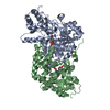
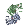
 Transaminase
  Keywords
 Function and homology information
 Authors
 Citation
 Structure visualization
 Downloads & links
 Other downloads
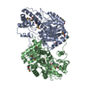


 PDBj
PDBj Assembly
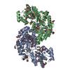


 Mass: 22.990 Da / Num. of mol.: 2 / Source method: obtained synthetically / Formula: Na
 Mass: 35.453 Da / Num. of mol.: 2 / Source method: obtained synthetically / Formula: Cl
 Mass: 62.068 Da / Num. of mol.: 19 / Source method: obtained synthetically / Formula: C2H6O2
 Mass: 354.436 Da / Num. of mol.: 2 / Source method: obtained synthetically / Formula: C16H34O8 / Comment: precipitant*YM
 Sample preparation
 / Beamline: 23-ID-B / Wavelength: 0.94645, 0.97967
 Processing