 Movie
Movie Controller


+ Open data

- Basic information
| Entry | Database: PDB / ID: 6tor | ||||||
|---|---|---|---|---|---|---|---|
| Title | human O-phosphoethanolamine phospho-lyase | ||||||
 Components Components | Ethanolamine-phosphate phospho-lyase | ||||||
 Keywords Keywords | LYASE / O-phosphoethanolamine phospho-lyase / pyridoxal 5'-phosphate-dependent enzyme / phospholipid metabolism. | ||||||
| Function / homology |  Function and homology informationethanolamine-phosphate phospho-lyase / ethanolamine-phosphate phospho-lyase activity / Synthesis of PE / phosphatidylethanolamine biosynthetic process / transaminase activity / pyridoxal phosphate binding / mitochondrial matrix Function and homology informationethanolamine-phosphate phospho-lyase / ethanolamine-phosphate phospho-lyase activity / Synthesis of PE / phosphatidylethanolamine biosynthetic process / transaminase activity / pyridoxal phosphate binding / mitochondrial matrixSimilarity search - Function | ||||||
| Biological species |  Homo sapiens (human) Homo sapiens (human) | ||||||
| Method | X-RAY DIFFRACTION / SYNCHROTRON / MOLECULAR REPLACEMENT / Resolution: 2.05 Å | ||||||
 Authors Authors | Vettraino, C. / Donini, S. / Parisini, E. | ||||||
 Citation Citation | Journal: Acta Crystallogr.,Sect.F / Year: 2020 Title: Structural characterization of human O-phosphoethanolamine phospho-lyase. Authors: Vettraino, C. / Peracchi, A. / Donini, S. / Parisini, E. #1: Journal: Acta Crystallogr.,Sect.F / Year: 2020Title: Structural characterization of human O-phosphoethanolamine phospho-lyase. Authors: Vettraino, C. / Peracchi, S. / Donini, S. / Parisini, E. | ||||||
| History |
|
- Structure visualization
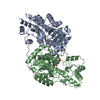
| Structure viewer | Molecule: MolmilJmol/JSmol |
|---|
- Downloads & links
-Download
| PDBx/mmCIF format | 6tor.cif.gz | 184.1 KB | Display | PDBx/mmCIF format |
|---|---|---|---|---|
| PDB format | pdb6tor.ent.gz | 143.5 KB | Display | PDB format |
| PDBx/mmJSON format | 6tor.json.gz | Tree view | PDBx/mmJSON format | |
| Others |  Other downloads Other downloads |
-Validation report
| Arichive directory | https://data.pdbj.org/pub/pdb/validation_reports/to/6torftp://data.pdbj.org/pub/pdb/validation_reports/to/6tor | HTTPS FTP |
|---|
-Related structure data
| Related structure data |  5g4iS S: Starting model for refinement |
|---|---|
| Similar structure data |


-Links
 PDBj
PDBj- Assembly
| Deposited unit | 
| ||||||||
|---|---|---|---|---|---|---|---|---|---|
| 1 |
| ||||||||
| Unit cell |
|
-Components
| #1: Protein | / Alanine--glyoxylate aminotransferase 2-like 1 Mass: 55757.750 Da / Num. of mol.: 2 Source method: isolated from a genetically manipulated source Source: (gene. exp.) Homo sapiens (human) / Gene: ETNPPL, AGXT2L1 / Production host:  Escherichia coli (E. coli) Escherichia coli (E. coli)References: UniProt: Q8TBG4, ethanolamine-phosphate phospho-lyase#2: Chemical | ChemComp-GOL / Glycerol  Mass: 92.094 Da / Num. of mol.: 6 / Source method: obtained synthetically / Formula: C3H8O3 Mass: 92.094 Da / Num. of mol.: 6 / Source method: obtained synthetically / Formula: C3H8O3#3: Chemical |   Mass: 248.173 Da / Num. of mol.: 2 / Source method: obtained synthetically / Formula: C8H13N2O5P / Feature type: SUBJECT OF INVESTIGATION Mass: 248.173 Da / Num. of mol.: 2 / Source method: obtained synthetically / Formula: C8H13N2O5P / Feature type: SUBJECT OF INVESTIGATION#4: Water | ChemComp-HOH / | Water Mass: 18.015 Da / Num. of mol.: 571 / Source method: isolated from a natural source / Formula: H2O Mass: 18.015 Da / Num. of mol.: 571 / Source method: isolated from a natural source / Formula: H2OHas ligand of interest | Y | |
|---|
-Experimental details
-Experiment
| Experiment | Method: X-RAY DIFFRACTION / Number of used crystals: 1 |
|---|
- Sample preparation
| Crystal | Density Matthews: 1.93 Å3/Da / Density % sol: 36.15 % |
|---|---|
| Crystal grow | Temperature: 298 K / Method: vapor diffusion, hanging drop / pH: 7 Details: 20% PEG 3350, 100 mM Bis Tris propane pH 7.0, 200 mM ammonium sulfate, and 3% MPD. |
-Data collection
| Diffraction | Mean temperature: 100 K / Serial crystal experiment: N |
|---|---|
| Diffraction source | Source: SYNCHROTRON / Site: SLS  / Beamline: X06DA / Wavelength: 1 Å / Beamline: X06DA / Wavelength: 1 Å |
| Detector | Type: PSI PILATUS 6M / Detector: PIXEL / Date: Oct 15, 2018 |
| Radiation | Protocol: SINGLE WAVELENGTH / Monochromatic (M) / Laue (L): M / Scattering type: x-ray |
| Radiation wavelength | Wavelength: 1 Å / Relative weight: 1 |
| Reflection | Resolution: 2.05→69.09 Å / Num. obs: 54318 / % possible obs: 99.99 % / Redundancy: 4.9 % / Biso Wilson estimate: 16.39 Å2 / CC1/2: 0.992 / Rpim(I) all: 0.068 / Net I/σ(I): 10.1 |
| Reflection shell | Resolution: 2.05→2.16 Å / Rmerge(I) obs: 0.102 / Num. unique obs: 54318 |
- Processing
| Software |
| ||||||||||||||||||||||||||||||||||||||||||||||||||||||||||||
|---|---|---|---|---|---|---|---|---|---|---|---|---|---|---|---|---|---|---|---|---|---|---|---|---|---|---|---|---|---|---|---|---|---|---|---|---|---|---|---|---|---|---|---|---|---|---|---|---|---|---|---|---|---|---|---|---|---|---|---|---|---|
| Refinement | Method to determine structure: MOLECULAR REPLACEMENT Starting model: 5G4I Resolution: 2.05→69.09 Å / Cor.coef. Fo:Fc: 0.955 / Cor.coef. Fo:Fc free: 0.94 / SU B: 4.186 / SU ML: 0.11 / Cross valid method: THROUGHOUT / σ(F): 0 / ESU R: 0.184 / ESU R Free: 0.152 / Stereochemistry target values: MAXIMUM LIKELIHOOD Details: HYDROGENS HAVE BEEN ADDED IN THE RIDING POSITIONS U VALUES : REFINED INDIVIDUALLY
| ||||||||||||||||||||||||||||||||||||||||||||||||||||||||||||
| Solvent computation | Ion probe radii: 0.8 Å / Shrinkage radii: 0.8 Å / VDW probe radii: 1.2 Å / Solvent model: MASK | ||||||||||||||||||||||||||||||||||||||||||||||||||||||||||||
| Displacement parameters | Biso max: 108.21 Å2 / Biso mean: 21 Å2 / Biso min: 4.75 Å2
| ||||||||||||||||||||||||||||||||||||||||||||||||||||||||||||
| Refinement step | Cycle: final / Resolution: 2.05→69.09 Å
| ||||||||||||||||||||||||||||||||||||||||||||||||||||||||||||
| Refine LS restraints |
| ||||||||||||||||||||||||||||||||||||||||||||||||||||||||||||
| LS refinement shell | Resolution: 2.05→2.103 Å / Rfactor Rfree error: 0 / Total num. of bins used: 20
|
