 Movie
Movie Controller

[English] 日本語
 Yorodumi
- PDB-3fvb: Crystal structure of ferritin (bacterioferritin) from Brucella me... -
+ Open data

- Basic information
| Entry | Database: PDB / ID: 3fvb | ||||||
|---|---|---|---|---|---|---|---|
| Title | Crystal structure of ferritin (bacterioferritin) from Brucella melitensis | ||||||
 Components Components | Bacterioferritin | ||||||
 Keywords Keywords | METAL BINDING PROTEIN / NIAID / SSGCID / deCODE / ferritin / Structural Genomics / Seattle Structural Genomics Center for Infectious Disease | ||||||
| Function / homology |  Function and homology informationferroxidase / ferroxidase activity / ferric iron binding / iron ion transport / intracellular iron ion homeostasis Function and homology informationferroxidase / ferroxidase activity / ferric iron binding / iron ion transport / intracellular iron ion homeostasisSimilarity search - Function | ||||||
| Biological species |  Brucella melitensis biovar Abortus 2308 (bacteria) Brucella melitensis biovar Abortus 2308 (bacteria) | ||||||
| Method | X-RAY DIFFRACTION / SYNCHROTRON / MOLECULAR REPLACEMENT / molecular replacement / Resolution: 1.806 Å | ||||||
 Authors Authors | Seattle Structural Genomics Center for Infectious Disease (SSGCID) | ||||||
 Citation Citation | Journal: To be Published Title: Crystal structure of ferritin from Brucella melitensis Authors: Seattle Structural Genomics Center for Infectious Disease (SSGCID) | ||||||
| History |
|
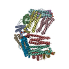
- Structure visualization
| Structure viewer | Molecule: MolmilJmol/JSmol |
|---|
- Downloads & links
-Download
| PDBx/mmCIF format | 3fvb.cif.gz | 93.7 KB | Display | PDBx/mmCIF format |
|---|---|---|---|---|
| PDB format | pdb3fvb.ent.gz | 68.4 KB | Display | PDB format |
| PDBx/mmJSON format | 3fvb.json.gz | Tree view | PDBx/mmJSON format | |
| Others |  Other downloads Other downloads |
-Validation report
| Arichive directory | https://data.pdbj.org/pub/pdb/validation_reports/fv/3fvbftp://data.pdbj.org/pub/pdb/validation_reports/fv/3fvb | HTTPS FTP |
|---|
-Related structure data
| Related structure data |  1jgcS S: Starting model for refinement |
|---|---|
| Similar structure data | |
| Other databases |


-Links
 PDBj
PDBj


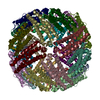
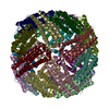
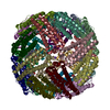
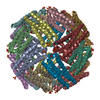
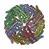
- Assembly
| Deposited unit | 
| |||||||||||||||||||||||||||
|---|---|---|---|---|---|---|---|---|---|---|---|---|---|---|---|---|---|---|---|---|---|---|---|---|---|---|---|---|
| 1 | x 12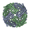
| |||||||||||||||||||||||||||
| Unit cell |
| |||||||||||||||||||||||||||
| Components on special symmetry positions |
|
-Components
-Protein , 1 types, 2 molecules AB
| #1: Protein | Mass: 20881.416 Da / Num. of mol.: 2 Source method: isolated from a genetically manipulated source Details: Expressed as a N-terminal hexahis tag with 3C protease cleavage site. Fusion tag was not cleaved prior to crystallization. Source: (gene. exp.) Brucella melitensis biovar Abortus 2308 (bacteria)Strain: biovar Abortus 2308 / Gene: bfr, BAB2_0675 / Plasmid: AVA0421 / Production host: Escherichia coli (E. coli) / References: UniProt: Q2YKI4 |
|---|
-Non-polymers , 7 types, 383 molecules 


| #2: Chemical | ChemComp-HEM / Heme B Mass: 616.487 Da / Num. of mol.: 1 / Source method: obtained synthetically / Formula: C34H32FeN4O4 Mass: 616.487 Da / Num. of mol.: 1 / Source method: obtained synthetically / Formula: C34H32FeN4O4 | ||||||||||
|---|---|---|---|---|---|---|---|---|---|---|---|
| #3: Chemical | Iron Mass: 55.845 Da / Num. of mol.: 3 / Source method: obtained synthetically / Formula: Fe Mass: 55.845 Da / Num. of mol.: 3 / Source method: obtained synthetically / Formula: Fe#4: Chemical | ChemComp-MG /  Mass: 24.305 Da / Num. of mol.: 6 / Source method: obtained synthetically / Formula: Mg Mass: 24.305 Da / Num. of mol.: 6 / Source method: obtained synthetically / Formula: Mg#5: Chemical | ChemComp-NA / |  Mass: 22.990 Da / Num. of mol.: 1 / Source method: obtained synthetically / Formula: Na Mass: 22.990 Da / Num. of mol.: 1 / Source method: obtained synthetically / Formula: Na#6: Chemical | Chloride Mass: 35.453 Da / Num. of mol.: 3 / Source method: obtained synthetically / Formula: Cl Mass: 35.453 Da / Num. of mol.: 3 / Source method: obtained synthetically / Formula: Cl#7: Chemical | Imidazole Mass: 69.085 Da / Num. of mol.: 2 / Source method: obtained synthetically / Formula: C3H5N2 Mass: 69.085 Da / Num. of mol.: 2 / Source method: obtained synthetically / Formula: C3H5N2#8: Water | ChemComp-HOH / | WaterMass: 18.015 Da / Num. of mol.: 367 / Source method: isolated from a natural source / Formula: H2O |
-Experimental details
-Experiment
| Experiment | Method: X-RAY DIFFRACTION / Number of used crystals: 1 |
|---|
- Sample preparation
| Crystal | Density Matthews: 2.66 Å3/Da / Density % sol: 53.84 % |
|---|---|
| Crystal grow | Temperature: 289 K / Method: vapor diffusion, sitting drop / pH: 7.5 Details: 12.4 mg/mL protein, 30% PEG 400, 0.2 M MGCl2, 0.1 M HEPES pH 7.5, 0.1 M imidazole; Crystal ID 200992h8. Original crystal hit was in JCSG+ D2, which was optimized using the Emerald Biosystems ...Details: 12.4 mg/mL protein, 30% PEG 400, 0.2 M MGCl2, 0.1 M HEPES pH 7.5, 0.1 M imidazole; Crystal ID 200992h8. Original crystal hit was in JCSG+ D2, which was optimized using the Emerald Biosystems ADDit Additive Screen. , VAPOR DIFFUSION, SITTING DROP, temperature 289K |
-Data collection
| Diffraction | Mean temperature: 100 K | ||||||||||||||||||||||||||||||||||||||||||||||||||||||||||||||||||
|---|---|---|---|---|---|---|---|---|---|---|---|---|---|---|---|---|---|---|---|---|---|---|---|---|---|---|---|---|---|---|---|---|---|---|---|---|---|---|---|---|---|---|---|---|---|---|---|---|---|---|---|---|---|---|---|---|---|---|---|---|---|---|---|---|---|---|---|
| Diffraction source | Source: SYNCHROTRON / Site: APS  / Beamline: 23-ID-D / Wavelength: 1.03322 Å / Beamline: 23-ID-D / Wavelength: 1.03322 Å | ||||||||||||||||||||||||||||||||||||||||||||||||||||||||||||||||||
| Detector | Type: MARMOSAIC 300 mm CCD / Detector: CCD / Date: Nov 5, 2008 | ||||||||||||||||||||||||||||||||||||||||||||||||||||||||||||||||||
| Radiation | Protocol: SINGLE WAVELENGTH / Monochromatic (M) / Laue (L): M / Scattering type: x-ray | ||||||||||||||||||||||||||||||||||||||||||||||||||||||||||||||||||
| Radiation wavelength | Wavelength: 1.03322 Å / Relative weight: 1 | ||||||||||||||||||||||||||||||||||||||||||||||||||||||||||||||||||
| Reflection | Resolution: 1.8→50 Å / Num. obs: 40564 / % possible obs: 100 % / Redundancy: 9.2 % / Rmerge(I) obs: 0.081 / Χ2: 1.151 / Net I/σ(I): 26.769 | ||||||||||||||||||||||||||||||||||||||||||||||||||||||||||||||||||
| Reflection shell |
|
-Phasing
| Phasing | Method: molecular replacement |
|---|
- Processing
| Software |
| |||||||||||||||||||||||||||||||||||||||||||||||||||||||||||||||||||||||||||||||||||||||||||||||||||||||||||||||||||||||||||||||||||||||||||||||||||
|---|---|---|---|---|---|---|---|---|---|---|---|---|---|---|---|---|---|---|---|---|---|---|---|---|---|---|---|---|---|---|---|---|---|---|---|---|---|---|---|---|---|---|---|---|---|---|---|---|---|---|---|---|---|---|---|---|---|---|---|---|---|---|---|---|---|---|---|---|---|---|---|---|---|---|---|---|---|---|---|---|---|---|---|---|---|---|---|---|---|---|---|---|---|---|---|---|---|---|---|---|---|---|---|---|---|---|---|---|---|---|---|---|---|---|---|---|---|---|---|---|---|---|---|---|---|---|---|---|---|---|---|---|---|---|---|---|---|---|---|---|---|---|---|---|---|---|---|---|
| Refinement | Method to determine structure: MOLECULAR REPLACEMENT Starting model: 1JGC, molecule A Resolution: 1.806→50 Å / Cor.coef. Fo:Fc: 0.961 / Cor.coef. Fo:Fc free: 0.948 / WRfactor Rfree: 0.21 / WRfactor Rwork: 0.176 / Occupancy max: 1 / Occupancy min: 0.33 / SU B: 2.149 / SU ML: 0.068 / Cross valid method: THROUGHOUT / σ(F): 0 / ESU R: 0.113 / ESU R Free: 0.111 / Stereochemistry target values: MAXIMUM LIKELIHOOD Details: HYDROGENS HAVE BEEN ADDED IN THE RIDING POSITIONS U VALUES : REFINED INDIVIDUALLY; CHLORIDE IONS MIGHT BE OTHER NEGATIVELY CHARGED IONS SUCH AS PHOSPHATE OR SULFATE, BUT CHLORIDE IONS ARE ...Details: HYDROGENS HAVE BEEN ADDED IN THE RIDING POSITIONS U VALUES : REFINED INDIVIDUALLY; CHLORIDE IONS MIGHT BE OTHER NEGATIVELY CHARGED IONS SUCH AS PHOSPHATE OR SULFATE, BUT CHLORIDE IONS ARE MOST LIKELY GIVEN CRYSTALLANT COMPOSITION
| |||||||||||||||||||||||||||||||||||||||||||||||||||||||||||||||||||||||||||||||||||||||||||||||||||||||||||||||||||||||||||||||||||||||||||||||||||
| Solvent computation | Ion probe radii: 0.8 Å / Shrinkage radii: 0.8 Å / VDW probe radii: 1.2 Å / Solvent model: MASK | |||||||||||||||||||||||||||||||||||||||||||||||||||||||||||||||||||||||||||||||||||||||||||||||||||||||||||||||||||||||||||||||||||||||||||||||||||
| Displacement parameters | Biso max: 65.17 Å2 / Biso mean: 25.375 Å2 / Biso min: 16.08 Å2 | |||||||||||||||||||||||||||||||||||||||||||||||||||||||||||||||||||||||||||||||||||||||||||||||||||||||||||||||||||||||||||||||||||||||||||||||||||
| Refinement step | Cycle: LAST / Resolution: 1.806→50 Å
| |||||||||||||||||||||||||||||||||||||||||||||||||||||||||||||||||||||||||||||||||||||||||||||||||||||||||||||||||||||||||||||||||||||||||||||||||||
| Refine LS restraints |
| |||||||||||||||||||||||||||||||||||||||||||||||||||||||||||||||||||||||||||||||||||||||||||||||||||||||||||||||||||||||||||||||||||||||||||||||||||
| LS refinement shell | Refine-ID: X-RAY DIFFRACTION / Total num. of bins used: 20
|
