 Movie
Movie Controller

[English] 日本語
 Yorodumi
- PDB-1jgc: The 2.6 A Structure Resolution of Rhodobacter capsulatus Bacterio... -
+ Open data

- Basic information
| Entry | Database: PDB / ID: 1jgc | ||||||
|---|---|---|---|---|---|---|---|
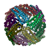
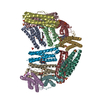
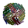
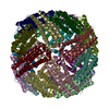
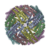
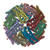
| Title | The 2.6 A Structure Resolution of Rhodobacter capsulatus Bacterioferritin with Metal-free Dinuclear Site and Heme Iron in a Crystallographic Special Position | ||||||
 Components Components | bacterioferritin | ||||||
 Keywords Keywords | METAL BINDING PROTEIN / Iron storage protein | ||||||
| Function / homology |  Function and homology informationferroxidase / ferroxidase activity / ferric iron binding / iron ion transport / intracellular iron ion homeostasis Function and homology informationferroxidase / ferroxidase activity / ferric iron binding / iron ion transport / intracellular iron ion homeostasisSimilarity search - Function | ||||||
| Biological species |  Rhodobacter capsulatus (bacteria) Rhodobacter capsulatus (bacteria) | ||||||
| Method | X-RAY DIFFRACTION / SYNCHROTRON / MIR / Resolution: 2.6 Å | ||||||
 Authors Authors | Cobessi, D. / Huang, L.-S. / Ban, M. / Pon, N.G. / Daldal, F. / Berry, E.A. | ||||||
 Citation Citation | Journal: Acta Crystallogr.,Sect.D / Year: 2002 Title: The 2.6 A resolution structure of Rhodobacter capsulatus bacterioferritin with metal-free dinuclear site and heme iron in a crystallographic 'special position'. Authors: Cobessi, D. / Huang, L.S. / Ban, M. / Pon, N.G. / Daldal, F. / Berry, E.A. | ||||||
| History |
|
- Structure visualization
| Structure viewer | Molecule: MolmilJmol/JSmol |
|---|
- Downloads & links
-Download
| PDBx/mmCIF format | 1jgc.cif.gz | 104.2 KB | Display | PDBx/mmCIF format |
|---|---|---|---|---|
| PDB format | pdb1jgc.ent.gz | 86.5 KB | Display | PDB format |
| PDBx/mmJSON format | 1jgc.json.gz | Tree view | PDBx/mmJSON format | |
| Others |  Other downloads Other downloads |
-Validation report
| Arichive directory | https://data.pdbj.org/pub/pdb/validation_reports/jg/1jgcftp://data.pdbj.org/pub/pdb/validation_reports/jg/1jgc | HTTPS FTP |
|---|
-Related structure data
| Similar structure data |
|---|


-Links
 PDBj
PDBj


- Assembly
| Deposited unit | 
| ||||||||
|---|---|---|---|---|---|---|---|---|---|
| 1 | x 8
| ||||||||
| Unit cell |
|
-Components
| #1: Protein | / BFR Mass: 18195.592 Da / Num. of mol.: 3 / Source method: isolated from a natural source / Source: (natural) Rhodobacter capsulatus (bacteria) / References: UniProt: Q59738#2: Chemical | Heme B  Mass: 616.487 Da / Num. of mol.: 3 / Source method: obtained synthetically / Formula: C34H32FeN4O4 Mass: 616.487 Da / Num. of mol.: 3 / Source method: obtained synthetically / Formula: C34H32FeN4O4#3: Water | ChemComp-HOH / | Water Mass: 18.015 Da / Num. of mol.: 21 / Source method: isolated from a natural source / Formula: H2O Mass: 18.015 Da / Num. of mol.: 21 / Source method: isolated from a natural source / Formula: H2O |
|---|
-Experimental details
-Experiment
| Experiment | Method: X-RAY DIFFRACTION / Number of used crystals: 1 |
|---|
- Sample preparation
| Crystal | Density Matthews: 3.11 Å3/Da / Density % sol: 60.4 % | ||||||||||||||||||||||||||||
|---|---|---|---|---|---|---|---|---|---|---|---|---|---|---|---|---|---|---|---|---|---|---|---|---|---|---|---|---|---|
| Crystal grow | Temperature: 293 K / Method: vapor diffusion / pH: 4.6 Details: octyl glucoside, NaOAc, PEK 4K, pH 4.6, VAPOR DIFFUSION, temperature 293K | ||||||||||||||||||||||||||||
| Crystal grow | *PLUS | ||||||||||||||||||||||||||||
| Components of the solutions | *PLUS
|
-Data collection
| Diffraction | Mean temperature: 100 K |
|---|---|
| Diffraction source | Source: SYNCHROTRON / Site: SSRL  / Beamline: BL7-1 / Wavelength: 1.08 Å / Beamline: BL7-1 / Wavelength: 1.08 Å |
| Detector | Type: MARRESEARCH / Detector: IMAGE PLATE / Date: Feb 17, 2000 / Details: MIRROR |
| Radiation | Monochromator: SI(111) / Protocol: SINGLE WAVELENGTH / Monochromatic (M) / Laue (L): M / Scattering type: x-ray |
| Radiation wavelength | Wavelength: 1.08 Å / Relative weight: 1 |
| Reflection | Resolution: 2.59→21.44 Å / Num. obs: 22679 / % possible obs: 99.4 % / Redundancy: 11.5 % / Rsym value: 0.077 / Net I/σ(I): 12.6 |
| Reflection shell | Resolution: 2.59→2.64 Å / Rsym value: 0.331 / % possible all: 94 |
| Reflection | *PLUS Highest resolution: 2.6 Å / Lowest resolution: 21.5 Å / Num. measured all: 260696 / Rmerge(I) obs: 0.077 |
| Reflection shell | *PLUS % possible obs: 94 % / Rmerge(I) obs: 0.331 |
- Processing
| Software |
| |||||||||||||||||||||||||
|---|---|---|---|---|---|---|---|---|---|---|---|---|---|---|---|---|---|---|---|---|---|---|---|---|---|---|
| Refinement | Method to determine structure: MIR / Resolution: 2.6→21.44 Å / σ(F): 0 / Stereochemistry target values: Engh & Huber
| |||||||||||||||||||||||||
| Displacement parameters | Biso mean: 27.2 Å2 | |||||||||||||||||||||||||
| Refine analyze |
| |||||||||||||||||||||||||
| Refinement step | Cycle: LAST / Resolution: 2.6→21.44 Å
| |||||||||||||||||||||||||
| Refine LS restraints |
| |||||||||||||||||||||||||
| LS refinement shell | Resolution: 2.6→2.66 Å
| |||||||||||||||||||||||||
| Software | *PLUS Name: CNS / Classification: refinement | |||||||||||||||||||||||||
| Refinement | *PLUS Lowest resolution: 21.5 Å / σ(F): 0 / % reflection Rfree: 4.9 % / Rfactor obs: 0.225 | |||||||||||||||||||||||||
| Solvent computation | *PLUS | |||||||||||||||||||||||||
| Displacement parameters | *PLUS Biso mean: 27.2 Å2 | |||||||||||||||||||||||||
| Refine LS restraints | *PLUS
| |||||||||||||||||||||||||
| LS refinement shell | *PLUS Rfactor Rfree: 0.398 / Rfactor obs: 0.314 |
