 Movie
Movie Controller


+ Open data

- Basic information
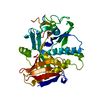
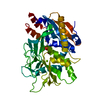
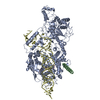
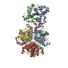
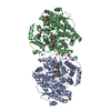
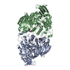
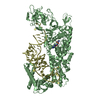
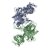
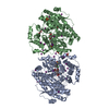
| Entry | Database: PDB / ID: 3fqd | ||||||
|---|---|---|---|---|---|---|---|
| Title | Crystal Structure of the S. pombe Rat1-Rai1 Complex | ||||||
 Components Components |
| ||||||
 Keywords Keywords | HYDROLASE/PROTEIN BINDING / Protein-Protein Complex /  Exonuclease / Hydrolase / mRNA processing / Nuclease / Nucleus / rRNA processing / Transcription / Transcription regulation / Transcription termination / Phosphoprotein / HYDROLASE-PROTEIN BINDING COMPLEX Exonuclease / Hydrolase / mRNA processing / Nuclease / Nucleus / rRNA processing / Transcription / Transcription regulation / Transcription termination / Phosphoprotein / HYDROLASE-PROTEIN BINDING COMPLEX | ||||||
| Function / homology |  Function and homology information Function and homology information5'-hydroxyl dinucleotide hydrolase activity / : / positive regulation of chromosome segregation / RNA NAD+-cap (NAD+-forming) hydrolase activity / Las1 complex / phosphodiesterase decapping endonuclease activity / mRNA 5'-diphosphatase activity / NAD-cap decapping / 5'-3' RNA exonuclease activity / nucleic acid metabolic process ...5'-hydroxyl dinucleotide hydrolase activity / : / positive regulation of chromosome segregation / RNA NAD+-cap (NAD+-forming) hydrolase activity / Las1 complex / phosphodiesterase decapping endonuclease activity / mRNA 5'-diphosphatase activity / NAD-cap decapping / 5'-3' RNA exonuclease activity / nucleic acid metabolic process / nuclear mRNA surveillance / nuclear-transcribed mRNA catabolic process / cleavage in ITS2 between 5.8S rRNA and LSU-rRNA of tricistronic rRNA transcript (SSU-rRNA, 5.8S rRNA, LSU-rRNA) / Hydrolases; Acting on acid anhydrides; In phosphorus-containing anhydrides / DNA-templated transcription termination / mRNA processing / GDP binding / Hydrolases; Acting on ester bonds; Exoribonucleases producing 5'-phosphomonoesters / RNA binding / metal ion binding / nucleus / cytosolSimilarity search - Function | ||||||
| Biological species |  Schizosaccharomyces pombe (fission yeast) Schizosaccharomyces pombe (fission yeast) | ||||||
| Method | X-RAY DIFFRACTION / SYNCHROTRON / Resolution: 2.2 Å | ||||||
 Authors Authors | Xiang, S. / Tong, L. | ||||||
 Citation Citation | Journal: Nature / Year: 2009 Title: Structure and function of the 5'-->3' exoribonuclease Rat1 and its activating partner Rai1. Authors: Xiang, S. / Cooper-Morgan, A. / Jiao, X. / Kiledjian, M. / Manley, J.L. / Tong, L. | ||||||
| History |
|
- Structure visualization
| Structure viewer | Molecule: MolmilJmol/JSmol |
|---|
- Downloads & links
-Download
| PDBx/mmCIF format | 3fqd.cif.gz | 437.6 KB | Display | PDBx/mmCIF format |
|---|---|---|---|---|
| PDB format | pdb3fqd.ent.gz | 367.8 KB | Display | PDB format |
| PDBx/mmJSON format | 3fqd.json.gz | Tree view | PDBx/mmJSON format | |
| Others |  Other downloads Other downloads |
-Validation report
| Arichive directory | https://data.pdbj.org/pub/pdb/validation_reports/fq/3fqdftp://data.pdbj.org/pub/pdb/validation_reports/fq/3fqd | HTTPS FTP |
|---|
-Related structure data


-Links
 PDBj
PDBj- Assembly
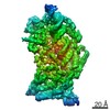
| Deposited unit | 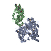
| ||||||||
|---|---|---|---|---|---|---|---|---|---|
| 1 |
| ||||||||
| Unit cell |
|
-Components
| #1: Protein | Mass: 103270.031 Da / Num. of mol.: 1 / Fragment: Rat1, residue 1-885 Source method: isolated from a genetically manipulated source Source: (gene. exp.) Schizosaccharomyces pombe (fission yeast)Gene: dhp1, Rat1, SPAC26A3.12c / Plasmid: pET24d / Production host:  Escherichia coli (E. coli) / Strain (production host): Rosetta (DE3) Escherichia coli (E. coli) / Strain (production host): Rosetta (DE3)References: UniProt: P40848, Hydrolases; Acting on ester bonds; Exoribonucleases producing 5'-phosphomonoesters |
|---|---|
| #2: Protein | Mass: 41315.492 Da / Num. of mol.: 1 Source method: isolated from a genetically manipulated source Source: (gene. exp.) Schizosaccharomyces pombe (fission yeast)Gene: din1, Rai1, SPAC19D5.06c / Plasmid: pET26b / Production host: Escherichia coli (E. coli) / Strain (production host): Rosetta (DE3) / References: UniProt: O13836 |
| #3: Chemical | ChemComp-MG /   Mass: 24.305 Da / Num. of mol.: 1 / Source method: obtained synthetically / Formula: Mg Mass: 24.305 Da / Num. of mol.: 1 / Source method: obtained synthetically / Formula: Mg |
| #4: Chemical | ChemComp-GOL / Glycerol  Mass: 92.094 Da / Num. of mol.: 1 / Source method: obtained synthetically / Formula: C3H8O3 Mass: 92.094 Da / Num. of mol.: 1 / Source method: obtained synthetically / Formula: C3H8O3 |
| #5: Water | ChemComp-HOH / Water Mass: 18.015 Da / Num. of mol.: 648 / Source method: isolated from a natural source / Formula: H2O Mass: 18.015 Da / Num. of mol.: 648 / Source method: isolated from a natural source / Formula: H2O |
-Experimental details
-Experiment
| Experiment | Method: X-RAY DIFFRACTION / Number of used crystals: 1 |
|---|
- Sample preparation
| Crystal | Density Matthews: 2.7 Å3/Da / Density % sol: 54.45 % |
|---|---|
| Crystal grow | Temperature: 295 K / Method: vapor diffusion, sitting drop / pH: 5 Details: 0.3 M sodium malonate (pH 5.0), 10 mM DTT and 14 % (w/v) PEG 3350, VAPOR DIFFUSION, SITTING DROP, temperature 295K |
-Data collection
| Diffraction | Mean temperature: 100 K | |||||||||||||||||||||||||||||||||||||||||||||||||||||||||||||||||||||||||||||
|---|---|---|---|---|---|---|---|---|---|---|---|---|---|---|---|---|---|---|---|---|---|---|---|---|---|---|---|---|---|---|---|---|---|---|---|---|---|---|---|---|---|---|---|---|---|---|---|---|---|---|---|---|---|---|---|---|---|---|---|---|---|---|---|---|---|---|---|---|---|---|---|---|---|---|---|---|---|---|
| Diffraction source | Source: SYNCHROTRON / Site: NSLS  / Beamline: X4C / Wavelength: 0.9806 Å / Beamline: X4C / Wavelength: 0.9806 Å | |||||||||||||||||||||||||||||||||||||||||||||||||||||||||||||||||||||||||||||
| Detector | Type: Mar m-165 CCD / Detector: CCD / Date: Feb 8, 2008 | |||||||||||||||||||||||||||||||||||||||||||||||||||||||||||||||||||||||||||||
| Radiation | Protocol: SINGLE WAVELENGTH / Monochromatic (M) / Laue (L): M / Scattering type: x-ray | |||||||||||||||||||||||||||||||||||||||||||||||||||||||||||||||||||||||||||||
| Radiation wavelength | Wavelength: 0.9806 Å / Relative weight: 1 | |||||||||||||||||||||||||||||||||||||||||||||||||||||||||||||||||||||||||||||
| Reflection | Resolution: 2.2→30 Å / Num. obs: 77326 / % possible obs: 92.5 % / Redundancy: 4 % / Rmerge(I) obs: 0.073 / Χ2: 1.088 / Net I/σ(I): 17.502 | |||||||||||||||||||||||||||||||||||||||||||||||||||||||||||||||||||||||||||||
| Reflection shell |
|
- Processing
| Software |
| ||||||||||||||||||||||||||||||||||||||||||||||||||||||||||||||||||||||||||||||||||||||||||
|---|---|---|---|---|---|---|---|---|---|---|---|---|---|---|---|---|---|---|---|---|---|---|---|---|---|---|---|---|---|---|---|---|---|---|---|---|---|---|---|---|---|---|---|---|---|---|---|---|---|---|---|---|---|---|---|---|---|---|---|---|---|---|---|---|---|---|---|---|---|---|---|---|---|---|---|---|---|---|---|---|---|---|---|---|---|---|---|---|---|---|---|
| Refinement | Resolution: 2.2→30 Å / Cor.coef. Fo:Fc: 0.939 / Cor.coef. Fo:Fc free: 0.915 / Occupancy max: 1 / Occupancy min: 0.5 / SU B: 11.018 / SU ML: 0.15 / Cross valid method: THROUGHOUT / σ(F): 0 / ESU R: 0.248 / ESU R Free: 0.208 / Stereochemistry target values: MAXIMUM LIKELIHOOD / Details: HYDROGENS HAVE BEEN ADDED IN THE RIDING POSITIONS
| ||||||||||||||||||||||||||||||||||||||||||||||||||||||||||||||||||||||||||||||||||||||||||
| Solvent computation | Ion probe radii: 0.8 Å / Shrinkage radii: 0.8 Å / VDW probe radii: 1.2 Å / Solvent model: MASK | ||||||||||||||||||||||||||||||||||||||||||||||||||||||||||||||||||||||||||||||||||||||||||
| Displacement parameters | Biso max: 163.38 Å2 / Biso mean: 44.537 Å2 / Biso min: 17.8 Å2
| ||||||||||||||||||||||||||||||||||||||||||||||||||||||||||||||||||||||||||||||||||||||||||
| Refinement step | Cycle: LAST / Resolution: 2.2→30 Å
| ||||||||||||||||||||||||||||||||||||||||||||||||||||||||||||||||||||||||||||||||||||||||||
| Refine LS restraints |
| ||||||||||||||||||||||||||||||||||||||||||||||||||||||||||||||||||||||||||||||||||||||||||
| LS refinement shell | Resolution: 2.2→2.257 Å / Total num. of bins used: 20
| ||||||||||||||||||||||||||||||||||||||||||||||||||||||||||||||||||||||||||||||||||||||||||
| Refinement TLS params. | Method: refined / Refine-ID: X-RAY DIFFRACTION
| ||||||||||||||||||||||||||||||||||||||||||||||||||||||||||||||||||||||||||||||||||||||||||
| Refinement TLS group |
|
