 Movie
Movie Controller

[English] 日本語
 Yorodumi
- PDB-3foj: Crystal Structure of SSP1007 From Staphylococcus saprophyticus su... -
+ Open data

- Basic information
| Entry | Database: PDB / ID: 3foj | ||||||
|---|---|---|---|---|---|---|---|
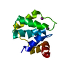
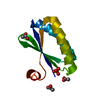
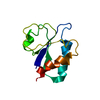
| Title | Crystal Structure of SSP1007 From Staphylococcus saprophyticus subsp. saprophyticus. Northeast Structural Genomics Target SyR101A. | ||||||
 Components Components | uncharacterized protein | ||||||
 Keywords Keywords |  structural genomics / unknown function / protein SSP1007 / PSI-2 / Protein Structure Initiative / Northeast Structural Genomics Consortium / NESG structural genomics / unknown function / protein SSP1007 / PSI-2 / Protein Structure Initiative / Northeast Structural Genomics Consortium / NESG | ||||||
| Function / homology |  Function and homology information Function and homology informationRhodanese-like domain / Oxidized Rhodanese; domain 1 / Rhodanese Homology Domain / Rhodanese-like domain / Rhodanese domain profile. / Rhodanese-like domain superfamily / Rhodanese-like domain / 3-Layer(aba) Sandwich / Alpha Beta Similarity search - Domain/homology | ||||||
| Biological species |  Staphylococcus saprophyticus subsp. saprophyticus ATCC 15305 (bacteria) Staphylococcus saprophyticus subsp. saprophyticus ATCC 15305 (bacteria) | ||||||
| Method | X-RAY DIFFRACTION / SYNCHROTRON / SAD / Resolution: 1.6 Å | ||||||
 Authors Authors | Seetharaman, J. / Abashidze, M. / Wang, H. / Janjua, H. / Foote, E.L. / Xiao, R. / Everett, J.K. / Acton, T.B. / Rost, B. / Montelione, G.T. ...Seetharaman, J. / Abashidze, M. / Wang, H. / Janjua, H. / Foote, E.L. / Xiao, R. / Everett, J.K. / Acton, T.B. / Rost, B. / Montelione, G.T. / Hunt, J.F. / Tong, L. / Northeast Structural Genomics Consortium (NESG) | ||||||
 Citation Citation | Journal: To be Published Title: Crystal Structure of SSP1007 From Staphylococcus saprophyticus subsp. saprophyticus. Northeast Structural Genomics Target SyR101A. Authors: Seetharaman, J. / Abashidze, M. / Wang, H. / Janjua, H. / Foote, E.L. / Xiao, R. / Everett, J.K. / Acton, T.B. / Rost, B. / Montelione, G.T. / Hunt, J.F. / Tong, L. | ||||||
| History |
|
- Structure visualization
| Structure viewer | Molecule: MolmilJmol/JSmol |
|---|
- Downloads & links
-Download
| PDBx/mmCIF format | 3foj.cif.gz | 30.4 KB | Display | PDBx/mmCIF format |
|---|---|---|---|---|
| PDB format | pdb3foj.ent.gz | 22.7 KB | Display | PDB format |
| PDBx/mmJSON format | 3foj.json.gz | Tree view | PDBx/mmJSON format | |
| Others |  Other downloads Other downloads |
-Validation report
| Arichive directory | https://data.pdbj.org/pub/pdb/validation_reports/fo/3fojftp://data.pdbj.org/pub/pdb/validation_reports/fo/3foj | HTTPS FTP |
|---|
-Related structure data
| Similar structure data | |
|---|---|
| Other databases |


-Links
 PDBj
PDBj- Assembly
| Deposited unit | 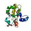
| ||||||||
|---|---|---|---|---|---|---|---|---|---|
| 1 |
| ||||||||
| Unit cell |
| ||||||||
| Details | monomer |
-Components
| #1: Protein | Mass: 11172.685 Da / Num. of mol.: 1 Source method: isolated from a genetically manipulated source Source: (gene. exp.) Staphylococcus saprophyticus subsp. saprophyticus ATCC 15305 (bacteria)Gene: SSP1007 / Production host: Escherichia coli (E. coli) / References: UniProt: Q49YI7 |
|---|---|
| #2: Chemical | ChemComp-NA /   Mass: 22.990 Da / Num. of mol.: 1 / Source method: obtained synthetically / Formula: Na Mass: 22.990 Da / Num. of mol.: 1 / Source method: obtained synthetically / Formula: Na |
| #3: Water | ChemComp-HOH / Water Mass: 18.015 Da / Num. of mol.: 123 / Source method: isolated from a natural source / Formula: H2O Mass: 18.015 Da / Num. of mol.: 123 / Source method: isolated from a natural source / Formula: H2O |
-Experimental details
-Experiment
| Experiment | Method: X-RAY DIFFRACTION / Number of used crystals: 1 |
|---|
- Sample preparation
| Crystal | Density Matthews: 1.7 Å3/Da / Density % sol: 27.77 % |
|---|---|
| Crystal grow | Temperature: 293 K / Method: vapor diffusion, sitting drop / pH: 5 Details: 150mM MgSO4, 100 MM Na3 Citrate, 20% PEG3350, VAPOR DIFFUSION, SITTING DROP, temperature 293K |
-Data collection
| Diffraction | Mean temperature: 100 K |
|---|---|
| Diffraction source | Source: SYNCHROTRON / Site: NSLS  / Beamline: X4A / Wavelength: 0.979 Å / Beamline: X4A / Wavelength: 0.979 Å |
| Detector | Type: ADSC QUANTUM 4 / Detector: CCD / Date: Nov 28, 2008 |
| Radiation | Protocol: SINGLE WAVELENGTH / Monochromatic (M) / Laue (L): M / Scattering type: x-ray |
| Radiation wavelength | Wavelength: 0.979 Å / Relative weight: 1 |
| Reflection | Resolution: 1.6→50 Å / Num. obs: 19428 / % possible obs: 99.7 % / Observed criterion σ(F): 0 / Observed criterion σ(I): 0 / Redundancy: 6.1 % / Biso Wilson estimate: 18.4 Å2 / Rmerge(I) obs: 0.076 / Net I/σ(I): 17.2 |
| Reflection shell | Resolution: 1.6→1.66 Å / Redundancy: 6 % / Mean I/σ(I) obs: 16 / Num. unique all: 1907 / Rsym value: 0.36 / % possible all: 99.4 |
- Processing
| Software |
| ||||||||||||||||||||||||||||||||||||
|---|---|---|---|---|---|---|---|---|---|---|---|---|---|---|---|---|---|---|---|---|---|---|---|---|---|---|---|---|---|---|---|---|---|---|---|---|---|
| Refinement | Method to determine structure: SAD / Resolution: 1.6→27.21 Å / Rfactor Rfree error: 0.008 / Data cutoff high absF: 119624.42 / Data cutoff low absF: 0 / Isotropic thermal model: RESTRAINED / Cross valid method: THROUGHOUT / σ(F): 0 / Stereochemistry target values: Engh & Huber / Details: BULK SOLVENT MODEL USED
| ||||||||||||||||||||||||||||||||||||
| Solvent computation | Solvent model: FLAT MODEL / Bsol: 42.7996 Å2 / ksol: 0.4 e/Å3 | ||||||||||||||||||||||||||||||||||||
| Displacement parameters | Biso mean: 18.1 Å2
| ||||||||||||||||||||||||||||||||||||
| Refine analyze |
| ||||||||||||||||||||||||||||||||||||
| Refinement step | Cycle: LAST / Resolution: 1.6→27.21 Å
| ||||||||||||||||||||||||||||||||||||
| Refine LS restraints |
| ||||||||||||||||||||||||||||||||||||
| LS refinement shell | Resolution: 1.6→1.69 Å / Total num. of bins used: 6
| ||||||||||||||||||||||||||||||||||||
| Xplor file |
|
